5544
Combining regional homogeneity and Meta-analysis to improve preoperative language mapping with resting-state functional MRI1Department of Imaging Physics, The University of Texas MD Anderson Cancer Center, Houston, TX, United States, 2Graduate Institute of Biomedical Electronics and Bioinformatics, National Taiwan University, Taipei, Taiwan, 3Department of Diagnostic Radiology, The University of Texas MD Anderson Cancer Center, Houston, TX, United States, 4Section of Neuropsychology, Department of Neuro-Oncology, The University of Texas MD Anderson Cancer Center, Houston, TX, United States, 5Department of Neurosurgery, The University of Texas MD Anderson Cancer Center, Houston, TX, United States
Synopsis
Resting-state (rs) fMRI has been shown its potential for pre-surgical mapping. Seed-correlation analysis is a commonly used approach for network detection. However, lesion-related spatial distortions and functional reorganization make the seed selection difficult for rs-fMRI mapping based on anatomical landmark alone. Here we proposed a novel approach to guide the seed selection for rs-fMRI mapping in patients with brain tumors by incorporating regional homogeneity (RH) confined by results of meta-analysis (MA). Our results showed performance that was equivalent to the seed localization guided by task-fMRI activation, suggesting the potential of RH+MA approach for rs-fMRI mapping in the clinical practice.
INTRODUCTION
Resting-state (rs) fMRI has been shown its helpfulness as a pre-operative mapping tool in localizing intrinsic functional networks during rest 1. One of the commonly used approaches of network detection is the seed-correlation analysis that imposes the prior knowledge of seed selection 2,3. However, lesion-related spatial distortions and functional reorganization make the seed selection difficult for mapping functional networks on the basis of anatomical landmark alone. In this study, we proposed a novel method to guide the seed selection for mapping the rs-fMRI language network in patients with brain tumors by incorporating data-driven regional homogeneity (RH) analysis confined by functional anatomy based on meta-analysis (MA).METHODS
The fMRI datasets from five patients with brain tumors were acquired using a gradient-echo EPI sequence (TR/TE/FA=2000 ms/25 ms/90°, voxel size=3.75×3.75×4 mm3) on a 3T clinical scanner. Language task-fMRI included a letter fluency (LETT) paradigm and a category fluency (CAT) paradigm. A total of 130 and 180 volumes was obtained from each of two task-fMRI datasets, and rs-fMRI dataset, respectively. The task-fMRI datasets were processed by AFNI including motion correction, realignment, de-spiking, de-trending, 4-mm FWHM spatial smoothing, and GLM analysis. Task-fMRI activation map was determined by setting a t threshold and a cluster-size threshold, corresponding to corrected p < 0.05. The rs-fMRI dataset was preprocessed as following steps: slice timing, motion correction, realignment, de-spiking, de-trending, regressing out covariates that included six motion parameters and fluctuations averaged over two masks of white matter and cerebrospinal fluid, band-pass filtering of 0.01–0.08 Hz, and 4-mm FWHM spatial smoothing. The RH map was calculated before spatial smoothing and confined within a mask obtained from inverse normalized MA map. The MA map was downloaded from Neurosynth using the term “language” that resulted from 885 studies 4 and constrained the results within the language regions-of-interest (ROI). This ROI encompasses bilateral middle frontal gyrus, bilateral inferior frontal gyrus, bilateral angular gyrus, bilateral supramarginal gyrus, and bilateral superior temporal gyrus, implemented by using the LONI Probabilistic Brain Atlas 5. The rs-fMRI mapping was calculated with a 6-mm spherical seed centering on a peak value of RH+MA and task-fMRI activation map within language ROI distant from the tumor. For quantitative comparison, Dice coefficient between the rs-fMRI maps and union of two task-fMRI maps were performed. Thresholds of rs-fMRI maps were optimized for maximizing the Dice coefficient, starting at a z value of 0.4 with a 0.02 increment. The Dice coefficient between two rs-fMRI methods for guiding seed selection were compared by using a Wilcoxon signed-rank test.RESULTS AND DISCUSSION
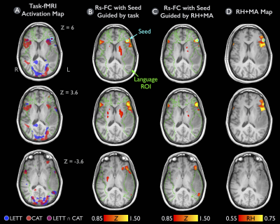
Among the 5 patients, 3 had lesions near Wernicke’s area and 2 near Broca’s area. Table 1 lists the resulted Dice coefficient for each patient determined from the whole brain and the language ROI. In the whole brain, Dice coefficient was on average 0.244 ± 0.084 and 0.238 ± 0.087 for rs-fMRI guided by task-fMRI and that by RH+MA map, respectively. These findings are in line with that of Branco at al.6, who reported the overlay between rs-fMRI and task-fMRI was on average 0.248. In the language ROI, the Dice coefficient was 0.315 ± 0.078 with rs-fMRI guided by task-fMRI map, and 0.291 ± 0.093 with rs-fMRI guided by RH+MA map. No significant difference (p = 0.63 and 0.31) were found in Dice coefficient between two rs-fMRI methods for both whole brain and language ROI. Figure 1A demonstrates that significant activations (p < 0.05, corrected) were detected in bilateral frontal areas for both task paradigms, left temporal/parietal area for LETT, and right temporal/parietal area for CAT. The RH+MA map showed scattered hot spots in the frontal regions (Fig. 1D). The rs-fMRI with use of either seeding approaches was helpful in detecting a clear language network close to the tumor, near temporal/parietal areas (Fig. 1B-C).CONCLUSION
This study proposed the RH+MA approach to guide the seed selection in rs-fMRI mapping. This new method showed no significant difference in Dice coefficient comparing to the seed selection based on task-fMRI activations. Our results suggest that the proposed method may be an effective and beneficial approach for mapping rs-fMRI functional network in the clinical practice, especially when a patient has difficulties in task compliance.
Acknowledgements
No acknowledgement found.References
1. Lang S, Duncan N, Northoff G. Resting-state functional magnetic resonance imaging: review of neurosurgical applications. Neurosurgery. 2014;74(5):453–64–discussion464–5. doi:10.1227/NEU.0000000000000307.
2. Biswal B, Yetkin FZ, Haughton VM, Hyde JS. Functional connectivity in the motor cortex of resting human brain using echo-planar MRI. Magn Reson Med. 1995;34(4):537–541.
3. Shimony JS, Zhang D, Johnston JM, Fox MD, Roy A, Leuthardt EC. Resting-state spontaneous fluctuations in brain activity: a new paradigm for presurgical planning using fMRI. Acad Radiol. 2009;16(5):578–583. doi:10.1016/j.acra.2009.02.001.
4. Yarkoni T, Poldrack RA, Nichols TE, Van Essen DC, Wager TD. Large-scale automated synthesis of human functional neuroimaging data. Nat Methods. 2011;8(8):665–670. doi:10.1038/nmeth.1635.
5. Shattuck DW, Mirza M, Adisetiyo V, et al. Construction of a 3D probabilistic atlas of human cortical structures. Neuroimage. 2008;39(3):1064–1080. doi:10.1016/j.neuroimage.2007.09.031.
6. Branco P, Seixas D, Deprez S, et al. Resting-State Functional Magnetic Resonance Imaging for Language Preoperative Planning. Front Hum Neurosci. 2016;10:11. doi:10.3389/fnhum.2016.00011.
Figures

