5543
Dynamic network analysis reveals altered temporal variability in brain regions after brain stroke: A longitudinal resting-state fMRI study1Department of Medical Imaging, Jinling Hospital, Nanjing Clinical School, Southern Medical University, Nanjing, China, Nanjing, China, 2Department of Radiology,The First Affiliated Hospital, Fujian Medical University, Fuzhou, China., Fuzhou, China, 3Department of Neurology, Jinling Hospital, Nanjing University School of Medicine, Nanjing, China, Nanjing, China, 4Department of Medical Imaging, Jinling Hospital, Nanjing University School of Medicine, Nanjing, China, Nanjing, China
Synopsis
In this work, we seek to investigate the longitudinal alteration of temporal variability in
resting-state brain network after cerebral stroke, by using a novel dynamic network analysis. Our study
illustrated a time dependent alteration of temporal variability in brain networks following stroke
recovery. These findings expand our understanding for dynamic properties of brain networks and
provide new insight into the underlying mechanisms of reorganization and integration of functional
networks over the recovery process after stroke.
Introduction
Resting-state functional magnetic resonance imaging (rs-fMRI) has proven to be a powerful tool for the assessment of neuroplasticity after stroke. Recent studies have revealed the dynamic nature of functional connectivity (FC) in brain network. Exploring time-varying FC networks offers a promising new approach to investigate the underlying mechanisms of reorganization and integration of functional networks over the recovery process after stroke. Here, using a novel dynamic network analysis based on rs-fMRI, we investigated the longitudinal alteration of temporal variability in brain regions after stroke.Methods
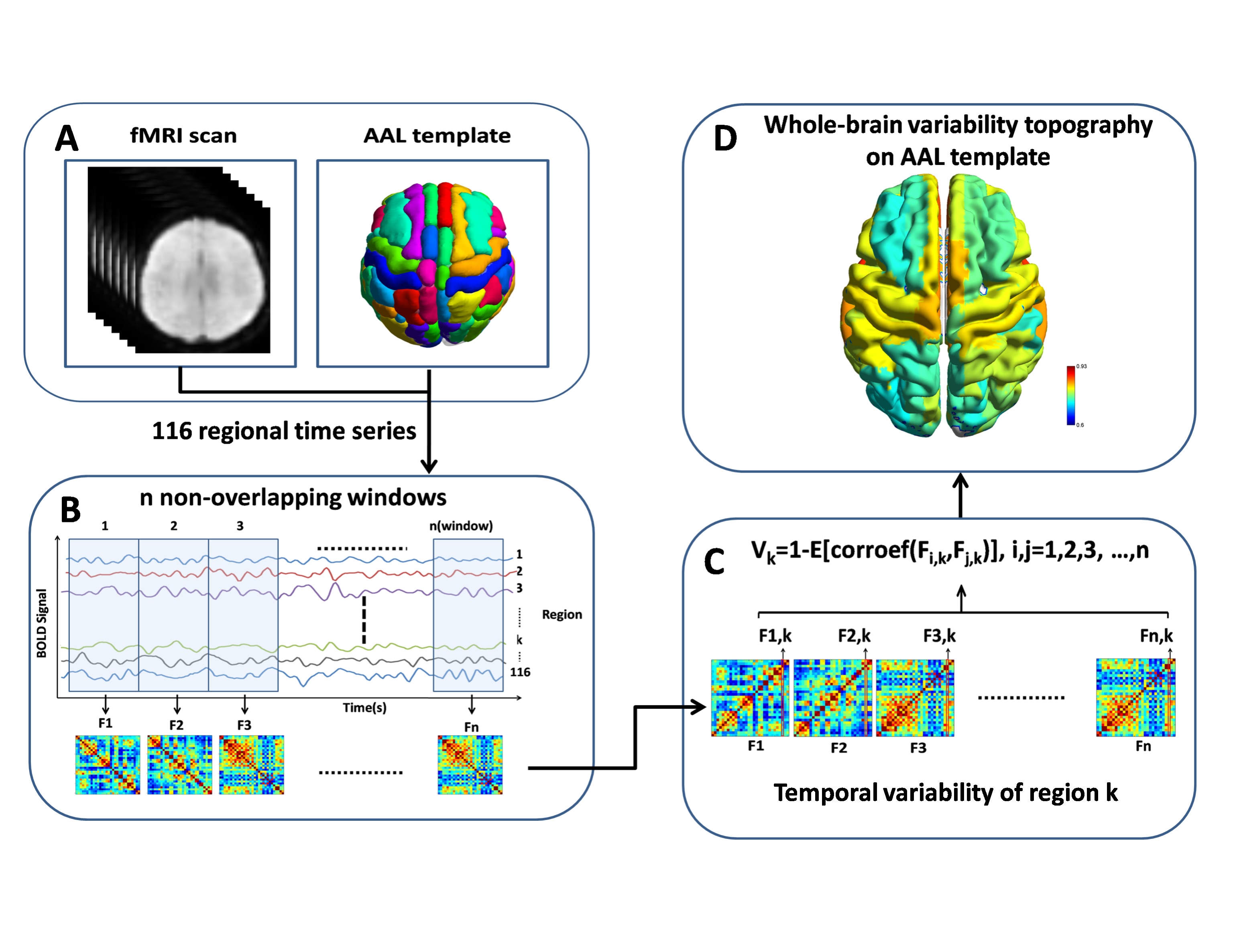
Nineteen stroke patients with motor impairment underwent rs-fMRI during the acute stage, the subacute stage and the early chronic stage. Nineteen age-matched healthy individuals were enrolled as baseline.Temporal variability in brain regions(AAL 116 template) was assessed based on dynamic functional network analysis described by zhang et al1 (Fig.1).Motor performance was evaluated with the Fugl-Meyer Assessment score. Temporal variability of brain networks between the groups and across the stages were compared by two-sample t-test and one way repeated measures ANOVA, respectively. The relationship between temporal variability in brain regions and clinical assessment was investigated by Pearson correlation analysis.
Results
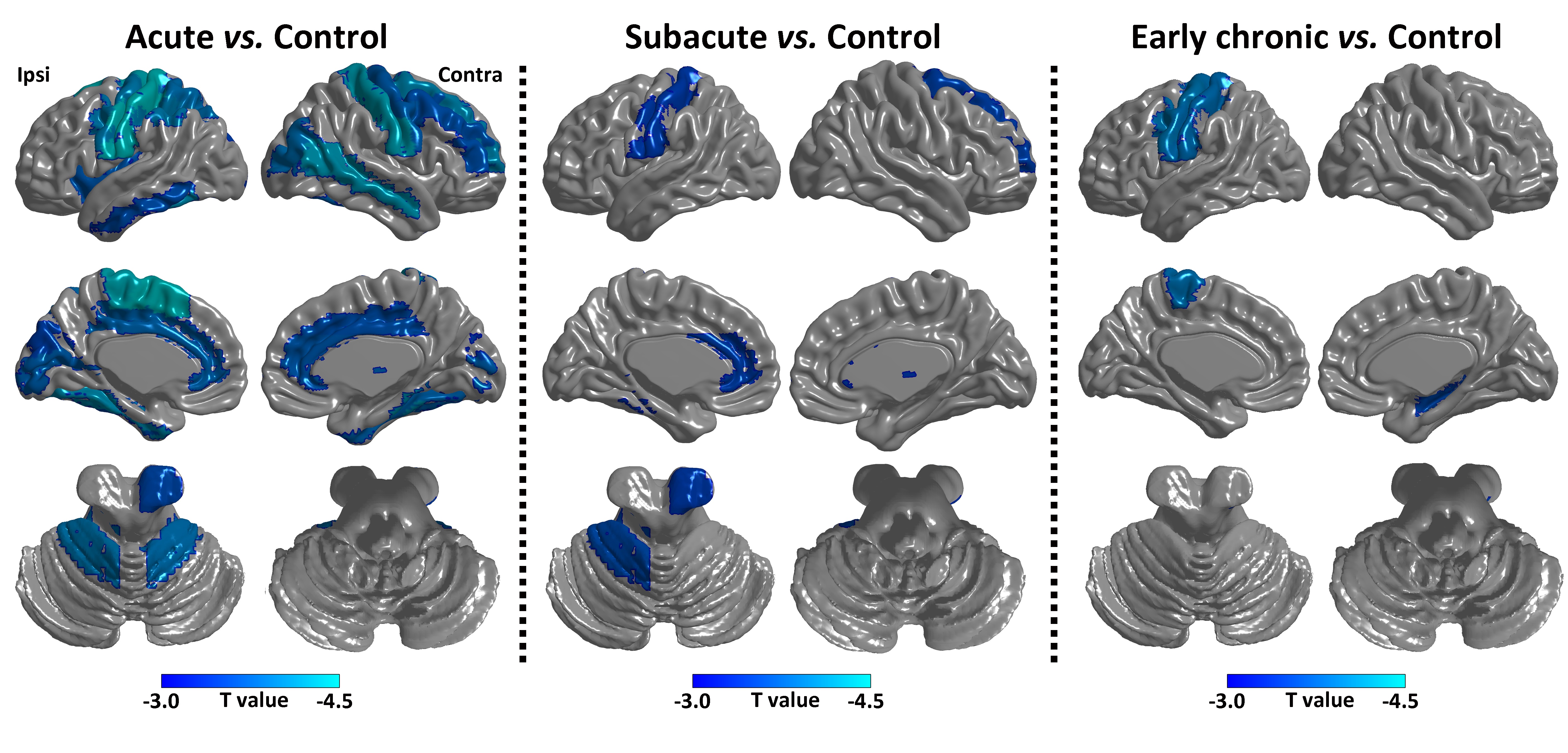
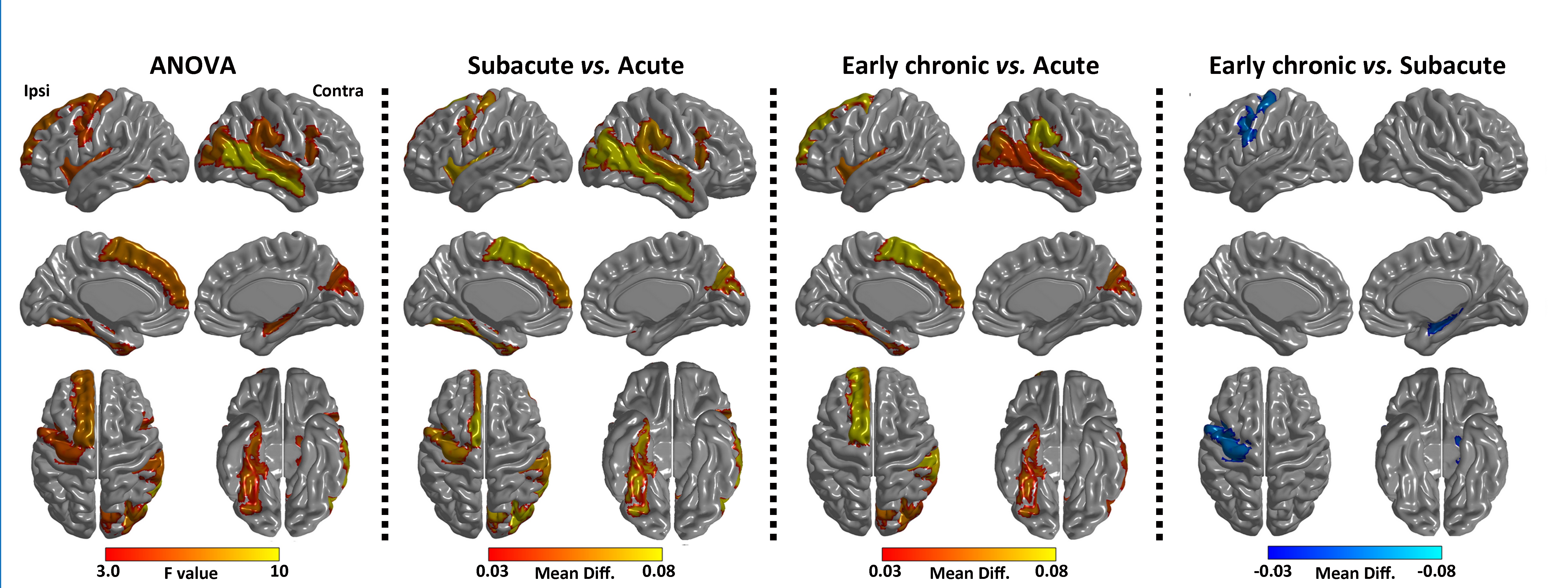
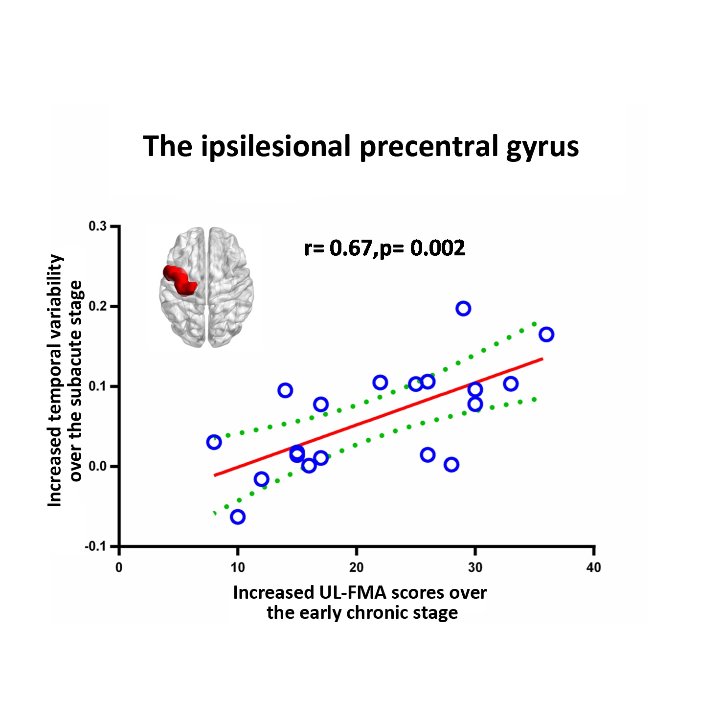
Compared with the healthy controls, stroke patients exhibited reduced temporal variability in all brain regions showing significant changes during the three stages. However, the number of these brain regions decreased over the stages of stroke progression. Except the ipsilesional sensorimotor cortex and the contralesional hippocampus, temporal variability in other regions was restored to the normal level during the early chronic stage. Compared to the acute stage, temporal variability in brain regions, including primary motor, auditory, visual cortices, significantly increased during the subacute stage. Most of these brain regions still kept increasing temporal variability during the early chronic stage, except the ipsilesional precentral gyrus (PreCG) and the contralesional hippocampus that exhibited reduced regional temporal variability when compared to the subacute stage. Increased temporal variability in the ipsilesional PreCG over the subacute stage was positively correlated with Motor recovery over the early chronic stage.Conclusions
Measuring temporal variability of brain networks may contribute to the understanding for the underlying mechanisms of reorganization and integration of functional networks over the recovery process after stroke, and can be severed as a complementary predictor for poststroke recovery.Acknowledgements
This study was supported by the special scientific research fund of public welfare profession of national health and family planning commission of China (201402019) and Jiangsu Province Foundation of China (BK20141373).References
1.Zhang, J., et al., Neural, electrophysiological and anatomical basis of brain-network variability and its characteristic changes in mental disorders. Brain, 2016.Figures