5531
Measurements of the Labeling Efficiency using super-selective Arterial Spin Labeling for Quantitative Flow Territory MappingThomas Lindner1, Mariya Krestina1, Olav Jansen1, and Michael Helle2
1Department of Radiology and Naeuroradiology, University Hospital Schleswig-Holstein, Kiel, Germany, 2Tomographic Imaging Department, Philips Research Laboratories, Hamburg, Germany
Synopsis
In this study, super-selective pseudo-continuous Arterial Spin Labeling was improved regarding the labeling efficiency and used in a volunteer study to evaluate the influence of the positioning of the focus with respect to the labeling efficiency.
Introduction
Quantifying Arterial Spin Labeling (ASL) perfusion allows for deeper insights into the physiology of the brain [1]. In selective ASL approaches used for flow territory mapping however, using assumptions of the labeling efficiency might result in inaccuracies. This becomes especially important in the case of super-selective ASL when the labeling focus is not positioned exactly at the artery of interest during scanning as the labeling efficiency reduces as a function of distance to the artery of interest [2]. Therefore, measuring the labeling efficiency of each scan seems important to obtain reliable results and to correct for potential miss-positioning errors. In this study, the labeling efficiency is measured using flow measurements of the tagged artery and taking into account the volume of the supplied flow territory [3]. The aim is to evaluate the labeling efficiency of super-selective ASL and considering these values when shifting the labeling spot step-by-step away from the artery of interest.Materials and Methods
In a first approach to optimize super-selective ASL, a fully randomized gradient pattern is implemented that reduces the labeling efficiency outside the artery of interest to near zero. Based on this tagging pattern, simulations were carried out to obtain the labeling efficiency values as function of distance to the tagged artery. These were validated in in-vivo experiments and the obtained values used for further processing of the data. All MR experiments were carried out using a Philips Achieva 3T scanner (Philips Healthcare, Best, The Netherlands). ASL parameters include: 1650ms Labeling duration and 1600ms post-labeling delay. Image acquisition was performed as 3D-GraSE scan with 2.3x2.3x5mm voxel size covering the whole brain. To evaluate the effect of misplacing the labeling spot, it was shifted by 2, 3, 5 and 9mm from the center of the artery of interest.Results and Discussion
MR experiments and simulations correspond well although the in-vivo experiments show a slight lower mean labeling efficiency across all offsets (Figure 1). The 2D simulation is shown in figure 2. Here also the drop of labeling efficiency to a value near zero outside the focus can be seen. Using the measured labeling efficiency value rather than the fixed one (0.85) shows that up to a distance of 3mm from the center of the artery, satisfying CBF values can still be obtained (Figure 3). At 5mm offset in neither case satisfying CBF values can be observed, which can be attributed to the already low SNR (in this case: amount of signal difference induced by labeling).Conclusion
Measuring the labeling efficiency in super-selective ASL experiments allows for a more accurate quantification of CBF values when the labeling focus exhibits an offset of up to 3mm from the center of the artery. The method of measuring the labeling efficiency is not limited to measuring the changes induced by shifting the labeling focus, but also flow velocities, which also influences labeling efficiency. Experiments on patients with different flow velocities are underway.Acknowledgements
No acknowledgement found.References
[1] Alsop D et. al. Magn Reson Med 2014;73:102-16 [2] Helle M et. al. Magn Reson Med 2010;64:777-86 [3] Juttukonda MR et. al. IEEE Biomedical Imaging 2016;1350-1353Figures
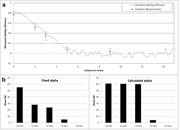
a) Simulation and validation
of the optimized super-selective labeling strategy. Inside, maximum labeling
efficiency can be obtained while outside the efficiency drops rapidly
b) Mean quantitative CBF
values of the flow territory (right internal carotid artery) using the fixed
value of labeling efficiency (α = 0.85), left and using the calculated values
of labeling efficiency, right.
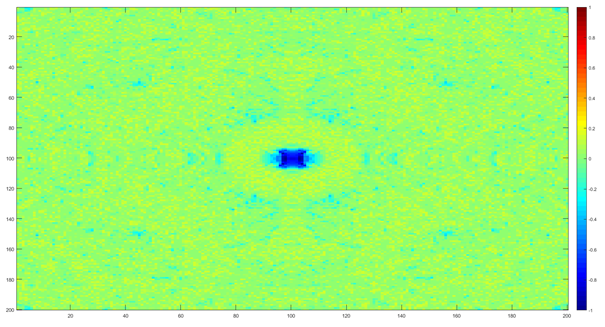
2D simulation of the Labeling efficiency showing high labeling efficiency inside the labeling focus and a reduction to near zero outside.
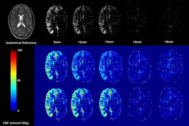
Relative SNR and
quantitative CBF values of the flow territory. The first row shows the relative
SNR of the flow territory with respect to the offset to the right internal
carotid artery. Up to 3mm offset, the territory stays clearly visible, while at
5mm it completely vanishes. The second row displays quantitative measures of
CBF using α = 0.85. The third row shows CBF maps using the calculated value for
α. Comparing the quantitative measures of 0, 2 and 3mm, it becomes evident that
the calculated α-value provides a more robust measure of CBF despite the shift
of the labeling focus.