5467
Predicting individual language task activation from resting state fMRI using a novel data-driven approachElizabeth Zakszewski1, Alexander Cohen1, Oiwi Parker Jones2, Saad Jbabdi2, and Yang Wang1
1Medical College of Wisconsin, Milwaukee, WI, United States, 2Oxford Centre for Functional Magnetic Resonance Imaging of the Brain (FMRIB), University of Oxford, Oxford, United Kingdom
Synopsis
This study is aimed to apply a newly developed machine learning approach to predict individual language network based on the resting state functional MRI (rs-fMRI). Despite the presence of significant variability of language network across subjects, the predicated language maps match excellently with the language task fMRI derived activation maps at the individual level. Our results suggest that rs-fMRI can be used as a promising clinical tool for mapping language network by using the novel processing approach.
Introduction
Functional MR (fMRI) imaging has been applied for preoperative mapping of eloquent brain regions related with important brain functions such as motor and language. Although task fMRI is successfully used in routine clinical care for operative management, resting state fMRI (rs-fMRI) has been investigated as an attractive alternative to task fMRI, specifically in patients who may be neurologically debilitated because of the presence of large brain lesions and may be unable to comply with a given task. However, only moderate group level rs-fMRI vs task fMRI language network concordance can be achieved by using existing methods.1-3 Most recently, a novel model based on machine learning (ML) has been proposed to accurately predict individual differences in brain activity.4 The method highlights a coupling between brain connectivity and function that can be captured at the level of individual subjects.4,5 Here, we take an in-depth look at the similarities between language task activations and activation maps predicted with the ML method.Methods
Total 30 healthy right-handed volunteers were scanned on a 3T MR scanner. For each subject, rs-fMRI was acquired using these parameters: TR/TE=2500/30ms, FA=90, voxel size=3.75x3.75x3mm3. The same subjects also performed a word generation task during a task fMRI acquisition using the same parameters as the rs-fMRI. Predictions of all subjects’ activation maps were made from rs-fMRI data using the methods of Parker Jones, et al.5 Briefly, all rs-fMRI data was preprocessed using the Human Connectome Project (HCP) minimal preprocessing pipeline6 and converted to the CIFTI format. Features were extracted from the combined rs-fMRI data using dual-step principle component analysis (PCA) followed by independent component analysis (ICA) to create a set of 70 group-level feature maps. These were combined with individual subjects’ rs-fMRI data to create a feature map for each subject. A ML algorithm was then trained to map from individual feature maps (predictors) to task activations. The beta coefficients were averaged with a leave-one-out analysis (leaving out that subject’s beta, creating a model that has not “seen” that subject) for each subject to create a predicted task activation map. This predicted activation map, the task-based activation map, and a map of the overlap between the two were viewed in the Connectome Workbench application. Additionally, individual language task activation maps were derived from a general linear model analysis on all task fMRI datasets.Results
Overall, predicated
activation maps generated from rs-fMRI data otline language networks in all
subjects. The correspondence between predicted language maps and task
activation maps match very well at the individual level (Fig1). Four example subjects are
shown in Figure 2. In all cases, significant overlap (green) between task and
predicted activation maps is seen in the left inferior frontal gyrus and
superior temporal gyrus. Lateralization is seen in that more activation occurs
in the left hemisphere (left) with less or none in the right hemisphere (right).Discussion and Conclusion
Despite the presence of significant variability of language network across subjects, the novel ML method of estimating language task activation based on rs-fMRI data is successful at predicting language areas at the individual level. Specifically, when thresholding to eliminate all but the strongest activations, the predicted maps provide an excellent match with the language task maps. Importantly, our results indicate clear language lateralization, as is expected for right-handed subjects. Our finding suggests that rs-fMRI can be used as a promising tool to provide important information on language network for neurosurgical planning and prognosis, particularly in pediatric population or those patients with issue of compliance.Acknowledgements
This work was supported by a Daniel M. Soref Charitable Trust Grant (to Y.W.).References
1. Sair HI, Yahyavi-Firouz-Abadi N, Calhoun VD, et al. Presurgical brain mapping of the language network in patients with brain tumors using resting-state fMRI: Comparison with task fMRI. Hum Brain Mapp. 2016;37(3):913-23. 2. Tie Y, Rigolo L, Norton IH, et al. Defining language networks from resting-state fMRI for surgical planning--a feasibility study. Hum Brain Mapp. 2014;35(3):1018-30. 3. Branco P, Sexias D, Deprez S, et al. Resting-State Functional Magnetic Resonance Imaging for Language Preoperative Planning. Front Hum Neurosci. 2016; doi: 10.3389/fnhum.2016.00011. 4. Tavor I, Parker-Jones O, Mars RB, et al. Task-free MRI predicts individual differences in brain activity during task performance. Science. 2016;352(6282):216-220. 5. Parker-Jones O, Voets NL, Adcock AE, et al. Resting connectivity predicts task activation in pre-surgical patients. Neuroimage – Clinical. 2017;13:378-385. 6. Glasser MF1, Sotiropoulos SN, Wilson JA, et al. WU-Minn HCP Consortium. The minimal preprocessing pipelines for the Human Connectome Project. Neuroimage. 2013;80:105-24.Figures
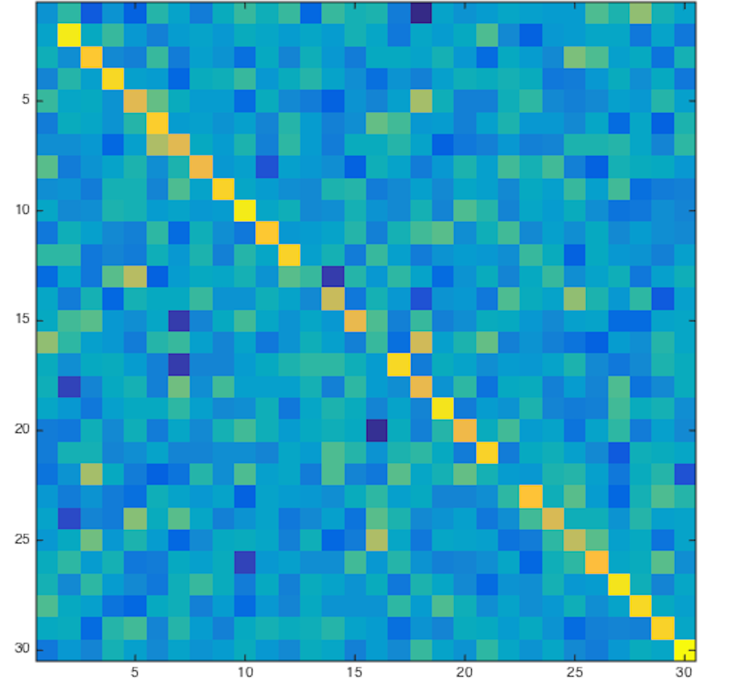
Figure1: Pearson’s
correlation coefficient matrices comparing all subjects’ predicted and actual
language task activation maps.
All matrices have been row- and column-normalized.
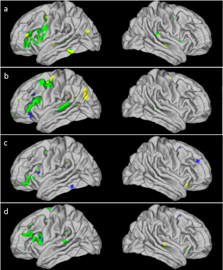
Figure2: Activation
maps for a word-generating task in 4 subjects (a-d). Overlaid are the actual
task activation map (yellow), the map predicted from the subject’s
resting-state scan (blue), and the overlap between the two (green). Activations
are seen in the inferior frontal gyrus (IFG) and superior temporal lobe (STL)
on the left, but not on the right, as is expected of a language-based task.