5461
Rapid 3D Blipped Spiral fMRI at 7 T1Institute for Biomedical Engineering, ETH Zurich and University of Zurich, Zurich, Switzerland, 2Translational Neuromodeling Unit, IBT, University of Zurich and ETH Zurich, Zurich, Switzerland, 3Skope Magnetic Resonance Technologies, Zurich, Switzerland
Synopsis
We present 3D blipped spiral fmri at 7T with a fast TR of 0.54s. Compared to 2D spirals, the 3D nature allows parallel imaging in z direction and blipped sampling of multiple planes that accelerate acquisition time, while the inherent averaging of multiple shots retains sufficient SNR. Intershot field inconsistency due to breathing and trajectory changes due to gradient heating in this high duty cycle sequence are compensated by NMR probe-based concurrent field monitoring, recovering high image quality and tSNR. We show the feasibility for fmri with 3D blipped spirals in a visual paradigm.
Introduction
Spiral imaging has been proposed as a promising acquisition technique for fMRI due to its high speed [1]. Recently, its successful deployment at 7T has been demonstrated for 2D sampling [2]. 3D imaging in comparison allows for parallel imaging in z direction and blipped sampling of multiple planes that accelerate acquisition time [3,4]. Compared to SMS acquisition [5], it also has a more benign SAR behavior due to simpler pulses, which becomes critical at ultra-high field and duty cycle. However, intershot inconsistencies e.g. due to breathing-related field changes, have stalled the adoption of the technique, as have unresolved calibration issues of the trajectories which change over time due to gradient heating for these high duty cycle sequences. Here, we show how these issues can be overcome by concurrent magnetic field monitoring and an expanded signal model incorporating static and dynamic offresonance and trajectory changes. We present 3D blipped spiral fMRI at 7T at a high temporal resolution (TR 540ms) with typical spatial resolution (3mm) and show its its feasibility in terms of tSNR and visual activation.Methods
Setup
Two healthy volunteers were scanned on a 7T Achieva system (Philips), using a 1-channel transmit, 32-channel receive coil (Nova Medical). 16 19F-NMR field probes [6] were placed in optimal positions between the receive and transmit module and connected to a dedicated MR acquisition system [7]. Visual stimulation was delivered via an LCD visor (VisuaStim).
Sequence and Trajectories
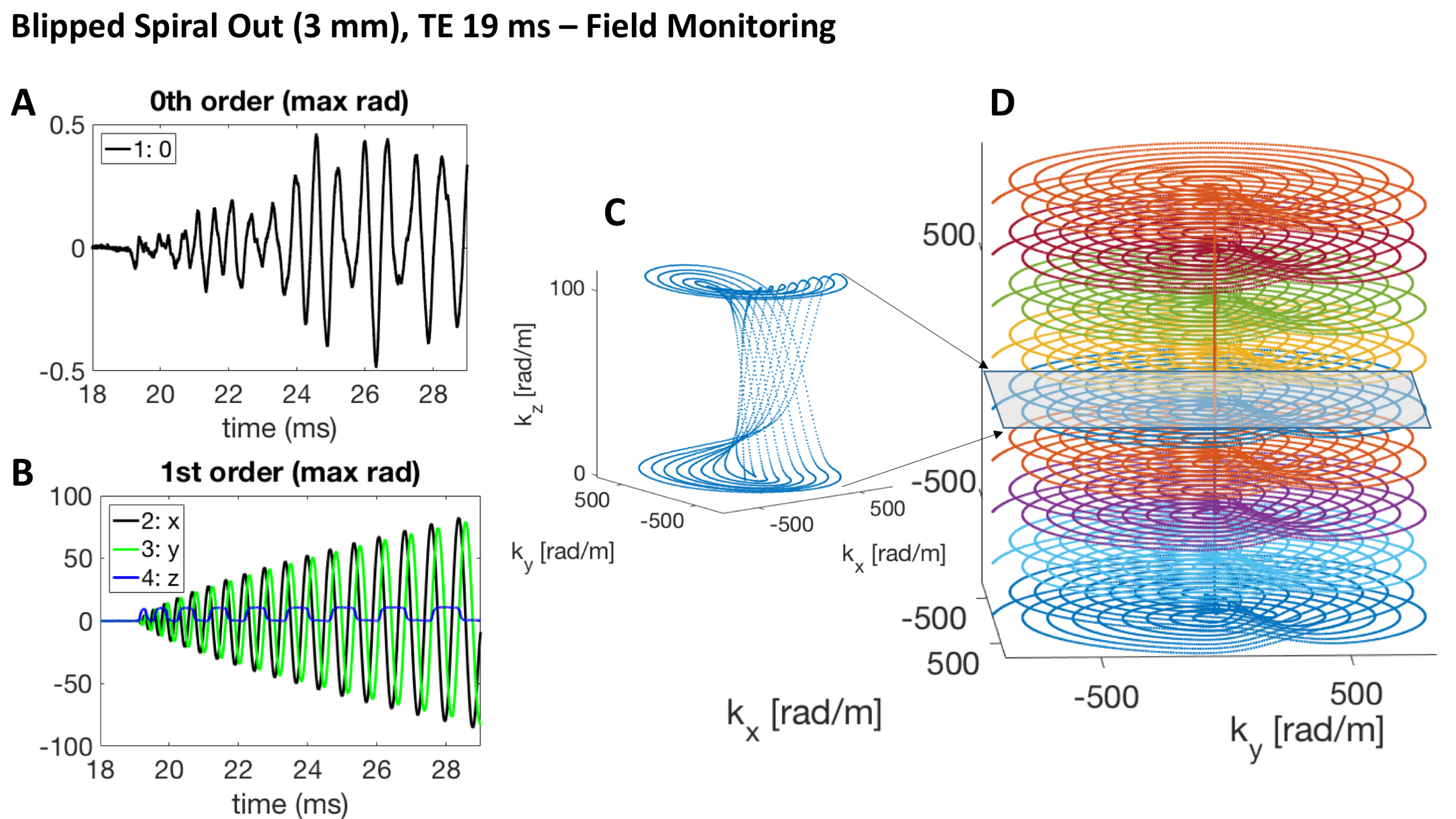
A 3D blipped spiral sequence with whole-brain coverage (FOV 230x230x120mm, 3 mm resolution) was designed with 9 interleaves (net 8-fold undersampling) covering 2 k-space planes each (12 ms readout duration, TE 19ms), amounting to a volume TR of 9*60 = 540 ms, exciting at the Ernst angle of 16 deg. The field probes were excited concurrently before the onset of each readout gradient.
Signal Processing and Image Reconstruction
For every time point during the imaging readout, 16 k-space coefficients – corresponding to a spherical harmonic basis set up to 3rd order – were computed from the probe signals. An iterative, CG-based image reconstruction was performed on the coil data (SENSE with multi-frequency interpolation, [9,10]). For the encoding model, the measured field dynamics up to first order (k0-k3) were used, in combination with the sensitivity- and static off-resonance maps [10, 11].
FMRI Analysis
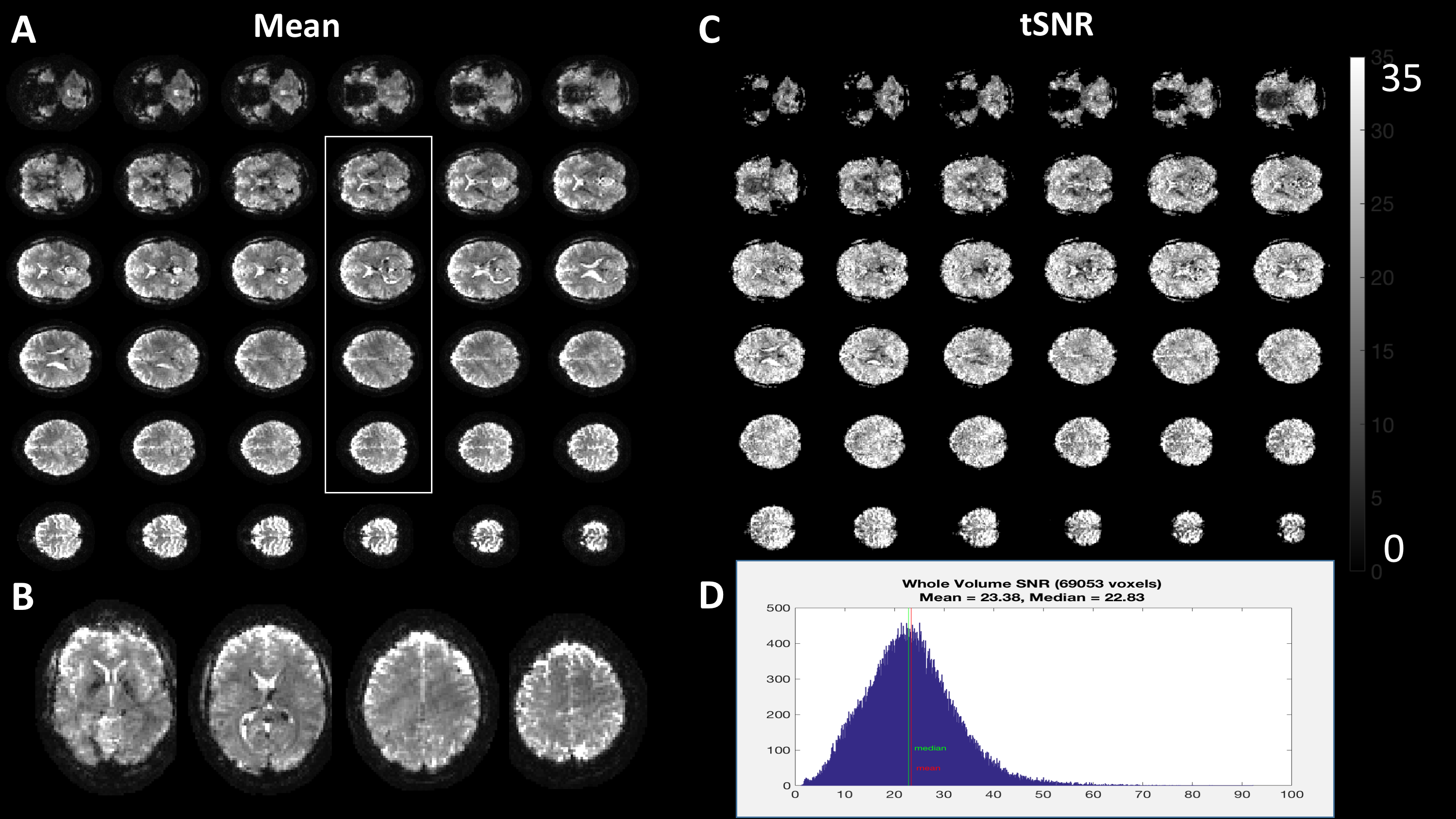
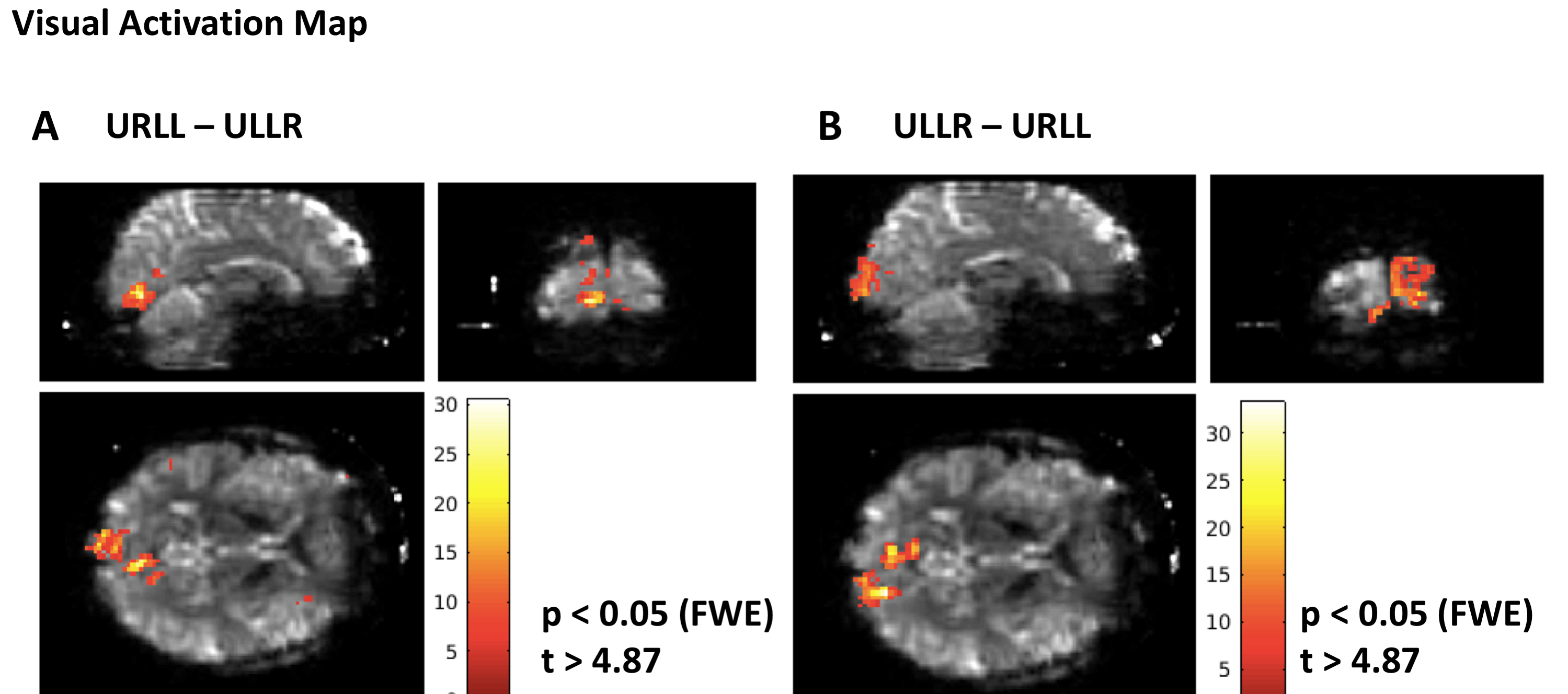
We evaluated tSNR in a time series of 100 volumes (resting state) and performed a visual quarter-field stimulation paradigm for 5 minutes (510 volumes). Preprocessing included realignment and smoothing (3mm). t-Contrasts are reported with whole-brain family-wise error correction.
Results
The trajectory could be successfully monitored with all interleaves and a very short re-excitation time of 60 ms for the NMR field probes (Fig. 1). Mean Image quality was good given the challenging conditions at high field and low spatial resolution. tSNR was sufficient given the short TR, between 20 and 30 (Fig. 2) with no spatial structure - indicating low G-factor noise enhancement. The successful detection of hemispherically distinct visual activation with a very conservative threshold (peak level family-wise error correction p=0.05) confirmed a sufficient sensitivity of this fast sequence for BOLD fMRI at 7T (Fig. 3).Discussion
We have shown that 3D blipped spiral fMRI is feasible at 7T. Its high acqusition efficiency can be successfully employed to accelerate the temporal resolution of fMRI to about 0.5s volume-TR for whole-brain coverage. 3D spirals may thus provide a viable alternative to multiband acquisitions at 7T due to their lower SAR. Furthermore, fast-TR fMRI opens up the possibility to study functional and effective brain connectivity with unprecedented precision due to the ability to sample BOLD effect transients and physiological noise sources.Acknowledgements
This work was supported by the NCCR “Neural Plasticity and Repair” at ETH Zurich and the University of Zurich (LK, KPP, KES), the René and Susanne Braginsky Foundation (KES), and the University of Zurich (KES). Technical support from Philips Healthcare, Best, The Netherlands, is gratefully acknowledged.References
[1] G.H. Glover, NeuroImage, 2012, 62, 706.
[2] Kasper et al., in Proc. Intl. Soc. Mag. Reson. Med. 25, 2017, 582
[3] M. Engel, L. Kasper, K.P. Pruessmann, in Proc. Intl. Soc. Mag. Reson. Med. 25, 2017, 5541.
[4] A. Stenger, C. Rettenmeier in Proc. Intl. Soc. Mag. Reson. Med. 25, 2017, 583
[5] K. Setsompop et al., Magnetic Resonance in Medicine, 2012, 67, 1210.
[6] N. De Zanche et al., Magnetic Resonance in Medicine, 2008, 60, 176―186.
[7] B.E. Dietrich et al., Magnetic Resonance in Medicine, 2016, 75, 1831.
[8] K.P. Pruessmann et al., Magnetic Resonance in Medicine, 2001, 46, 638―651.
[9] L.C. Man, J.M. Pauly, A. Macovski, Magnetic Resonance in Medicine, 1997, 37, 785.
[10] C. Barmet, N. De Zanche, K.P. Pruessmann, Magnetic Resonance in Medicine, 2008, 60, 187―197.
[11] B.J. Wilm et al., Magnetic Resonance in Medicine, 2011, 65, 1690.
Figures