5453
Enhanced BOLD Contrast-to-Noise-Ratio and Activation Sensitivity with the Intra-shot Adapted Keyhole (ISAK) Method1Biophysics, Medical College of Wisconsin, Milwaukee, WI, United States, 2Radiology, Medical College of Wisconsin, Milwaukee, WI, United States
Synopsis
A fast, multi-echo (ME) fMRI acquisition method, ISAK, is introduced by employing the classic keyhole method in a between-echo manner. ISAK acquires eight echoes within 70 ms, and the associated contrast-to-noise-ratio (CNR) improvement is up to more than 200%. The stronger blood-oxygen-level-dependent sensitivity, compared with regular ME fMRI with four echoes and standard single-echo fMRI without any acceleration, proves the benefit of trading a fraction of spatial samples in each image for more temporal echoes images in improving CNR.
Purpose
Multi-echo (ME) fMRI1 is increasingly recognized and accepted in acquiring brain activation signals for its capability to improve the BOLD contrast-to-noise ratio (CNR). However, within a limited data acquisition time, whether to acquire high temporal resolution in the time repetition (TR) domain or more echoes in the time-echo (TE) domain, in terms of the CNR, is still controversial. It is also challenging to acquire more echo images while maintaining a high spatial resolution for each image. To address these issues, we created the intra-shot adapted keyhole (ISAK) method and evaluated its effects on BOLD CNR and statistical power in fMRI studies.
Theory
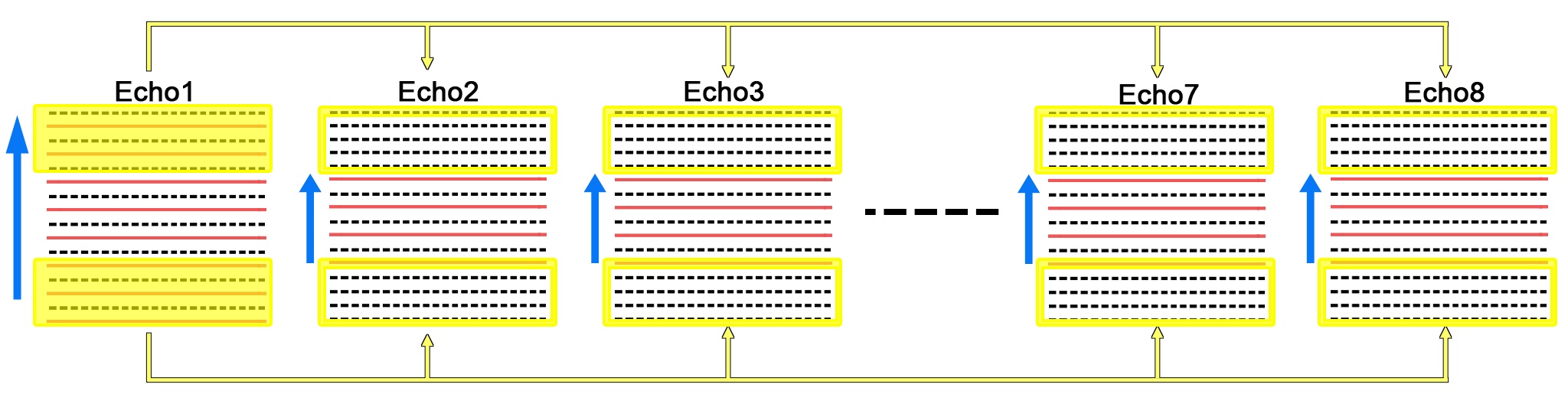
The keyhole method2 is classic in imaging acceleration across repetitions in that it samples only a central fraction of k-space (low k-space data), whereas the missing peripheral lines in the k-space (high k-space data) are supplied from the reference images acquired with full k-space data. We revitalized this method to accelerate ME acquisition in an across-echo manner. The data acquisition and reconstruction workflow are illustrated in Figure 1. A weighted substitution following equation 1 $$$S_{keyhole}=S_{full}\times exp^{\frac{TE_{full} - TE_{keyhole}}{T2^{*}}} (1)$$$ using peripheral samples from echo 1 for later echoes was carried out to avoid strip artifacts from the signal intensity discontinuity between echoes. The ISAK method not only preserves the advantage of acquiring contrast in the low-frequency data with high temporal resolution, it also maintains the changes in high-frequency data by acquiring a full k-space reference image within each repetition.Data Acquisition and Processing
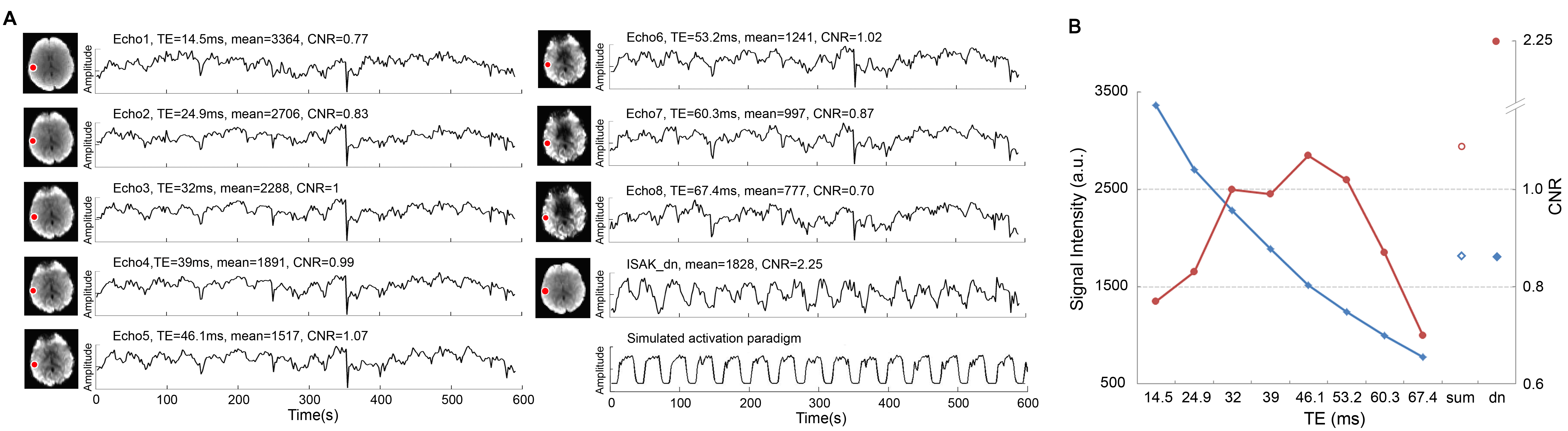
To quantify the CNR improvement, whole-brain scans with block design auditory stimulation were performed on 15 subjects by three methods: ISAK (eight TEs: 14.5, 24.9, 32.0, 39.0, 46.1, 53.2, 60.3, 67.4 ms), regular ME (four TEs: 14.5, 29.0, 43.4, 57.8 ms), and standard single echo (SE, TE=30 ms). The in-plane resolution was 3.75 mm isotropic across three methods, and the in-plane SENSE factor for ISAK and ME is 2. The number of slices and slice thickness are 35 and 3.5 mm for SE, 25 and 5 mm for the two other methods. Both ISAK and ME weighted combined all echo images then denoised the combined image using ME-ICA3, and SE was denoised by deconvolving white matter (WM), cerebral spinal fluid (CSF), and motion regressors. A general linear regression model was performed to calculate the CNR using SPM software4.Results
The ISAK method significantly increased voxelwise BOLD CNR after the summation of all echoes and denoising. As shown in Figure 2, the CNR acquired with the ISAK method improved more than 200% in comparison with that acquired with the SE method. It is worth noting that the obvious signal artifact at the time of 354 s across all eight echo images was successfully removed by the ISAK method.
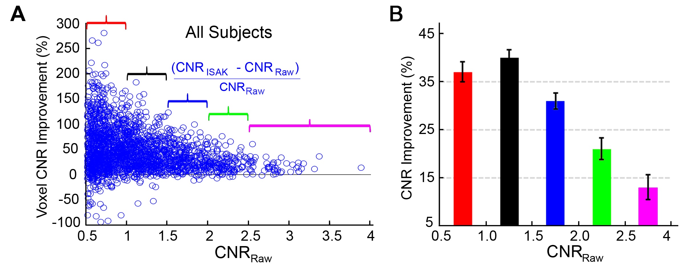
The ISAK method improves CNR in a CNRRaw-dependent manner, as shown in Figure 3. When the maximum CNR among the eight echo images, labeled as CNRRaw, is low, the percent improvement of CNR will be high. For example, when the mean voxelwise CNRRaw is smaller than 1, the mean CNR improvement in ISAK is 37%, whereas when the CNRRaw is more than 2.5, the mean CNR improvement is only 13%. This feature demonstrates the value of the ISAK method by showing that it is capable of restoring the CNR, especially when the CNR of the raw echo signal is poor (less than 1).
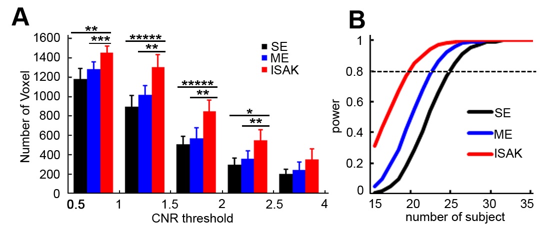
The BOLD CNR improvement acquired with the ISAK method has significantly increased BOLD signal detection sensitivity in comparison with conventional ME and SE acquisition methods. Figure 4A shows results from the paired two-sample t-test; the number of activated voxels was statistically more by the ISAK method, resulting in the strongest statistical power in detecting BOLD activation.
Discussion
The ISAK method can accelerate ME fMRI acquisition and increase the number of echo images, while maintaining the spatial resolution of each echo image. The outstanding performance in extracting BOLD contrast from noisy fMRI time series has significantly improved BOLD detection sensitivity and proves the benefit of statistical power in fMRI studies.Acknowledgements
No acknowledgement found.References
- S. Posse et al., Enhancement of BOLD-contrast sensitivity by single-shot multi-echo functional MR imaging. Magnetic resonance in medicine : official journal of the Society of Magnetic Resonance in Medicine / Society of Magnetic Resonance in Medicine 42, 87-97 (1999).
- J. J. van Vaals et al., "Keyhole" method for accelerating imaging of contrast agent uptake. Journal of magnetic resonance imaging : JMRI 3, 671-675 (1993).
- P. Kundu, S. J. Inati, J. W. Evans, W. M. Luh, P. A. Bandettini, Differentiating BOLD and non-BOLD signals in fMRI time series using multi-echo EPI. NeuroImage 60, 1759-1770 (2012).
- By members & collaborators of the Wellcome Department of Imaging Neuroscience: University College London, SPM12—Statistical Parametric Mapping, http://www.fil.ion.ucl.ac.uk/spm
Figures