5448
Multi-Band-Accelerated T2*-Weighted Inner-Field-of-View EPI of the Human Spinal Cord with Slice-Specific z-Shimming1Systems Neuroscience, Univ. Medical Center Hamburg-Eppendorf, Hamburg, Germany
Synopsis
T2*-weighted EPI suffers from severe signal dropouts and geometric distortions in the human spinal cord due to susceptibility differences between tissue, vertebrae, and vertebral bones. With 2D-selective RF (2DRF) excitations, the field-of-view can be focused to the spinal cord without aliasing which reduces geometric distortions, shortens echo times, and increases the signal-to-noise ratio significantly. Here, this approach is accelerated with simultaneous multi-slice (SMS) imaging to shorten the acquisition time. An individual temporal shift was applied to the different bands of a multi-band 2DRF envelope which corresponds to a slice- or band-specific z-shim that compensates for the specific through-slice dephasing effects.
Introduction
T2*-weighted echo-planar imaging is the standard imaging technique for BOLD-based functional neuroimaging. However, its application to the human spinal cord suffers from severe signal dropouts and geometric distortions due to the susceptibility differences between tissue, vertebrae, and vertebral bones (e.g. [1]), in particular if large fields-of-view are required to avoid aliasing artifacts [2]. With 2D-selective RF (2DRF) excitations [3,4], the field-of-view can be focused to the spinal cord without aliasing which has been shown to reduce geometric distortions, shorten echo times, and increase the signal-to-noise ratio significantly [2]. Here, this approach is accelerated with simultaneous multi-slice (SMS) imaging [5] to shorten the acquisition time. With an individual temporal shift for the different bands to be combined [6], a band- or slice specific z-shimming can be realized to minimize through-slice signal dephasingMethods
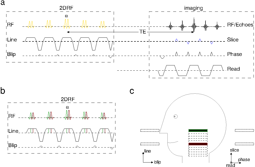
The basic pulse sequence and geometric setup are sketched in Fig. 1. Compared to a conventional EPI sequence, blipped-CAIPI gradient pulses were applied along the echo train [5] and the initial slice-selective RF excitation was replaced by a 2DRF excitation based on a fly-back blipped-planar trajectory (e.g. [7]) with the line and blip directions coinciding with the imaging slice and phase-encoding directions, respectively (Fig. 1a). The multi-band 2DRF envelope (yellow) is obtained by combining the single-band 2DRF envelopes after applying (i) a linear phase modulation to consider the different positions along the line direction and (ii) individual temporal shifts that correspond to the band-specific z-shim gradient moment required to compensate for through-slice dephasing [6] (Fig. 1b and c).
The desired 2DRF excitation profiles, either one (single-band) or two (multi-band) 4.0x32.0-mm2 (line x blip) rectangles (band distance between 40 and 75 mm), were realized with a trajectory resolution of 2.5x10.0 mm2 (line x blip) and a field-of-excitation of 210 mm to shift unwanted side excitations outside of the object for the target regions investigated (cf. Fig. 1c). Blipped-CAIPI gradient pulses were adjusted to an apparent shift of FOV/2 between the two bands of a multi-band 2DRF excitation.
Experiments were performed using a 3T whole-body MR system (Magnetom PrismaFit, Siemens, Germany) with a standard 64-channel head-neck coil on phantoms and healthy volunteers from which informed consent was obtained prior to the examination. The in-plane resolution was 1.0x1.0 mm2, the slice thickness between 4.0 and 5.0 mm. An inner FOV of 32 mm was used, 16 mm oversampling were added to account for the finite profile resolution in the blip/phase-encoding direction yielding a standard echo time of 30 ms. For 20 slices, repetition times (TR) of 1.550 s and 0.775 s could be achieved without and with SMS, respectively. Optimal z-shim gradient moments for each slice/band were determined from a single-band reference scan stepping through different setting for the z-shim.
Results
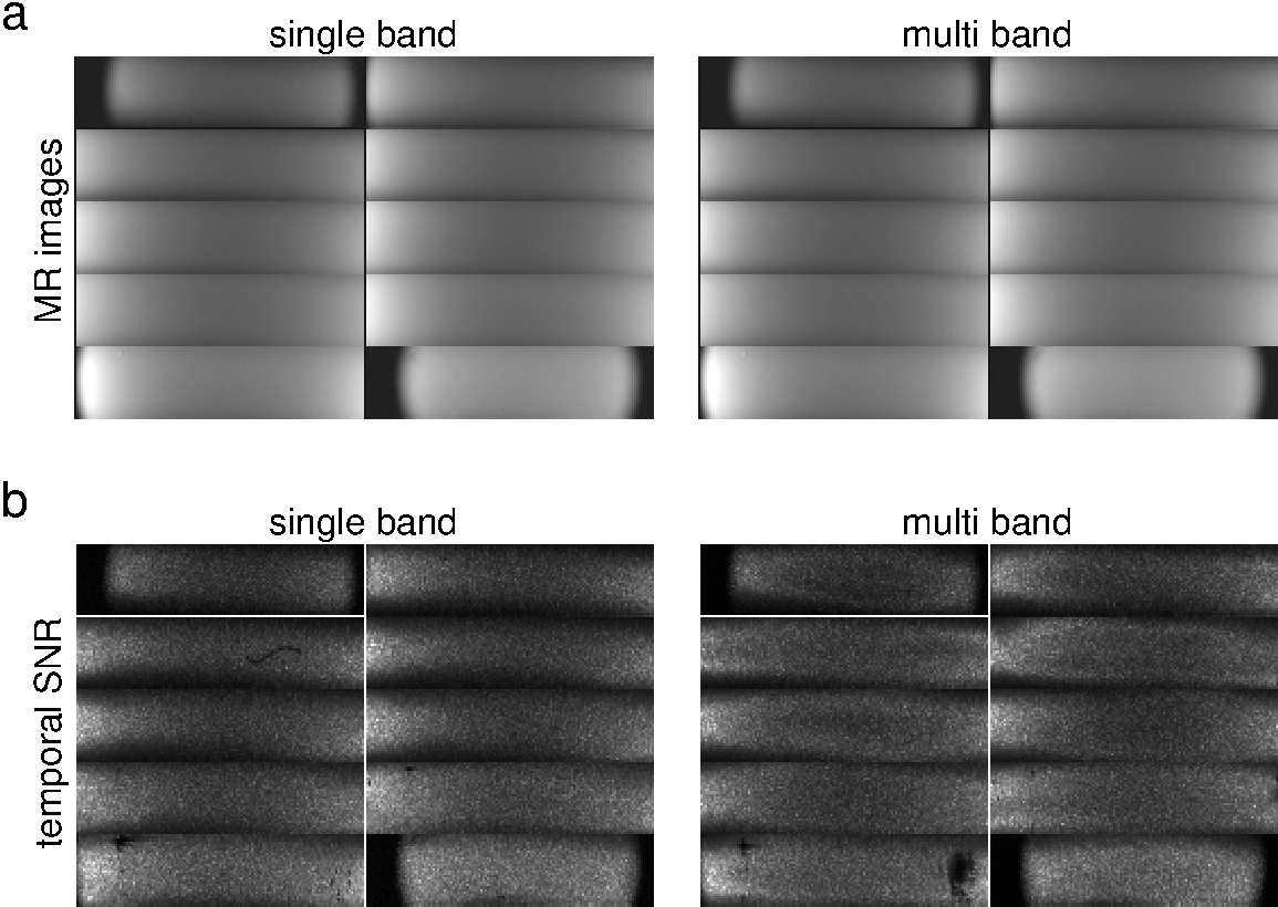
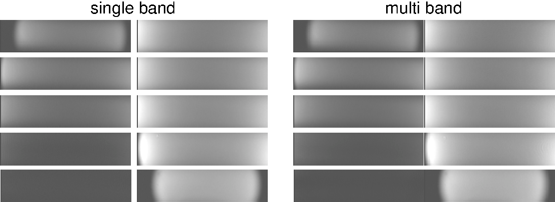
Results obtained in a water phantom for a TR of 6 s are summarized in Fig. 2 and 3. The image quality for standard and SMS acquisitions is very similar with no aliasing from unwanted side excitations, profile imperfections, or SMS (Fig. 2a). The temporal SNR calculated for 20 measurements (Fig. 2b) shows comparable results for single- and multi-band acquisitions. Outer slices tend to lose some SNR (between 5% and 10%) while inner slices gain it (up to 15%), most likely due to a non-uniform noise distribution of the SMS reconstruction algorithm. Figure 3 demonstrates that individual z-shim settings can be used for simultaneously excited bands without performance losses compared to conventional (single-band) excitations.
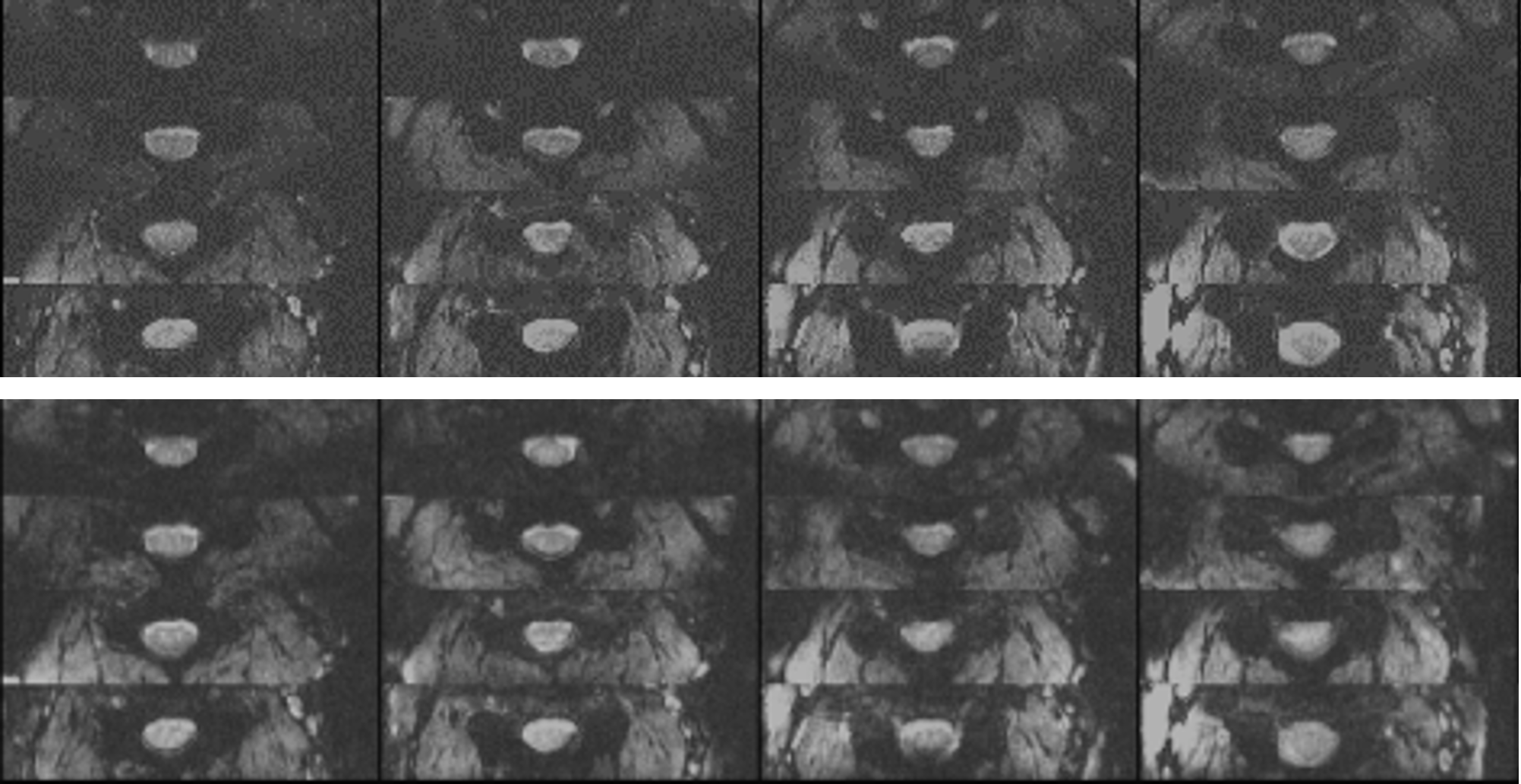
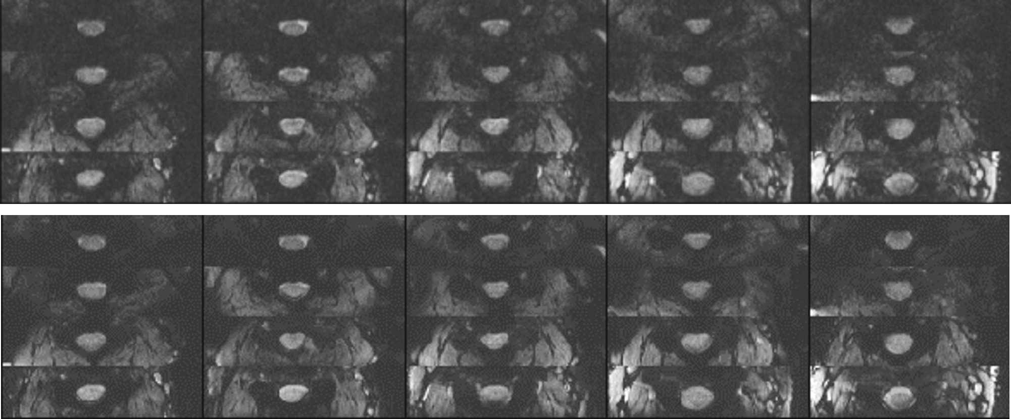
In vivo data acquired in the cervical section of the spinal cord are presented in Fig. 4 and 5. The signal gain with slice/band-specific z-shimming (data not shown) and the image quality achieved are similar for conventional and SMS acquisitions for an identical TR (1550 ms; Fig. 4). For the shorter TR achievable with multi-band acceleration (775 ms), the flip angle needs to be decreased and the SNR is reduced accordingly (Fig. 5). Nevertheless, the image quality obtained could be expected to be sufficient for BOLD-based functional imaging of the spinal cord.
Discussion
For cord sections below the cervical part, the field-of-excitation may have to be increased to ensure that the unwanted side excitations appear outside of the object. This will result in prolonged 2DRF excitations and echo times. Furthermore, the performance of the coils used needs to be evaluated to estimate the acceleration factors achievable.Conclusion
Simultaneous EPI of multiple inner fields-of-view with slice- or band-specific z-shimming are feasible and improve the temporal resolution of BOLD-based functional neuroimaging of the spinal cord. This could help to improve the detection and correction of motion and the modelling of physiological noise.Acknowledgements
Parts of this work were supported by Wings for Life.References
1. Finsterbusch J, Sprenger C, Büchel C. Single, slice-specific z-shim gradient pulses improve T2*-weighted imaging of the spinal cord. Neuroimage 2012; 59: 2307-2315.
2. Finsterbusch J. T2*-weighted echo-planar imaging of the spinal cord: full vs. inner fields-of-view. Proc. Int. Soc. Magn. Reson. Med. 25, 589 (2017).
3. Bottomley PA, Hardy CJ. Two-dimensional spatially selective spin inversion and spin-echo refocusing with a single nuclear magnetic resonance pulse. J. Appl. Phys. 1987; 62: 4284-4290.
4. Pauly J, Nishimura D, Macovski A. A k-space analysis of small-tip-angle excitations. J. Magn. Reson. 1989; 81: 43-56.
5. Setsompop K, Gagoski BA, Polimeni JR, Witzel T, Wedeen VJ, Wald LL. Blipped-controlled aliasing in parallel imaging for simultaneous multislice echo planar imaging with reduced g-factor penalty. Magn. Reson. Med. 2012; 67: 1210-1224.
6. Finsterbusch J. Multi-Band Slice-Selective and 2D-Selective RF Excitations with Band-Specific Dephasing Moments for Tailored z-Shimming. Proc. Int. Soc. Magn. Reson. Med. 24, 1881 (2016).
7. Finsterbusch J. Functional neuroimaging of inner fields-of-view with 2D-selective RF excitations. Magn. Reson. Imaging 2013; 31: 1228-35.
Figures