5438
Using Virtual Conjugate Coil reconstruction for statistical improvement in highly accelerated Simultaneous Multislice fMRI1Brain Imaging Centre, Research Centre for Natural Sciences, Hungarian Academy of Sciences, Budapest, Hungary, 2Department of Nuclear Techniques, Budapest University of Technology and Economics, Budapest, Hungary, 3Magnetic Resonance and X-ray Imaging Department, Fraunhofer Development Center X-ray Technology (EZRT), Würzburg, Germany, 4Athinoula A. Martinos Center for Biomedical Imaging, Charlestown, MA, United States, 5Department of Radiology, Harvard Medical School, Boston, MA, United States, 6Harvard-MIT Health Sciences and Technology, Massachusetts Institute of Technology, Cambridge, MA, United States, 7MR Application Predevelopment, Siemens Healthcare GmbH, Erlangen, Germany
Synopsis
Simultaneous multislice EPI is a popular acquisition method for high-temporal-resolution fMRI. However, at high acceleration factors, significant noise amplification could occur that can hinder the detection of activation. Using the Virtual Conjugate Coil concept, the g-factors of such measurements can be reduced. In this work we investigate the potential statistical improvement of using Virtual Conjugate Coil reconstruction in simultaneous multislice fMRI. Our results show that using Virtual Conjugate Coil reconstruction, first-level and group-level t-values, as well as first-level effect sizes and temporal signal-to-noise ratio are increased.
Introduction
Simultaneous multislice (SMS) EPI1-4 is an increasingly popular fMRI acquisition method due to its potential to significantly increase the temporal resolution, while avoiding the inherent $$$\sqrt{R}$$$ loss in signal-to-noise-ratio (SNR) of conventional parallel imaging. However, at high acceleration factors, significant noise amplification can still occur due to the numerical conditioning of the reconstruction (the so-called g-factor5), decreasing the ability to detect activations. Virtual Conjugate Coil6 (VCC) reconstruction is a phase-constrained method that can improve the SNR of SMS acquisitions7. It was recently shown8 that it can be combined with Partial Fourier (PF) imaging, making it suitable for fMRI where PF is commonly used to achieve BOLD-optimal TE. In this work, the benefit of VCC reconstruction on the fMRI statistical analysis is investigated for highly accelerated SMS EPI acquisitions.
Methods
Measurements were performed on a clinical 3T scanner (MAGNETOM Prisma, Siemens Healthcare, Erlangen, Germany), in accordance with institutional ethical regulations, with the informed consent of all volunteers. Image reconstruction and all calculations were performed offline using Matlab (The MathWorks, Natick, MA, USA) and SPM129. Ten healthy adults (mean±std age: 26.4±3.3 years, 6 females) were scanned while performing combined visual and motor tasks, similar as in Ref.10. Visual stimuli consisted of a 7.5-Hz flickering checkerboard stimulating one visual hemifield at a time. The motor task was a simple sequential finger-tapping. Volunteers were instructed to perform the tapping when the checkerboard was flickering, using the hand on the same side where the checkerboard appeared. The order of left and right task blocks was randomized with the stimulus duration fixed to 15.2 seconds. Resting intervals were randomly varied between 12.8 and 17.6 seconds, with 506.4 seconds total scanning time.
A prototype blipped-CAIPI SMS gradient-echo-EPI sequence1,11 was used for all functional measurements with 12-fold slice acceleration, using full-brain coverage with an isotropic 2 mm spatial resolution and a repetition time of 405 ms, without in-plane parallel imaging. Partial Fourier factor of 7/8 was used to achieve an echo time of 30 ms. Offline image reconstruction was performed using the Slice-GRAPPA algorithm1, with and without the inclusion of VCC signals to study the effect of VCC reconstruction on the following statistical analysis. Besides including or omitting VCC signals, all other reconstruction and processing parameters were identical.
fMRI preprocessing steps and statistical analysis were performed offline using SPM129. The data were realigned and normalized to MNI space, followed by a 3-mm isotropic Gaussian smoothing and a 128-second high-pass filter. First-level statistical analysis used a standard General Linear Model (GLM), with two regressors for right-side and left-side stimulation convolved by the canonical HRF9. Voxel-wise t-tests were performed to compare stimulus with baseline. Group-level random-effects analysis was also performed on the first-level effects. In addition to statistical analysis, temporal SNR (tSNR) maps were also computed to compare standard and VCC reconstructions, using the mean signal, and the standard deviation of the residual signal after the GLM fit.
Results
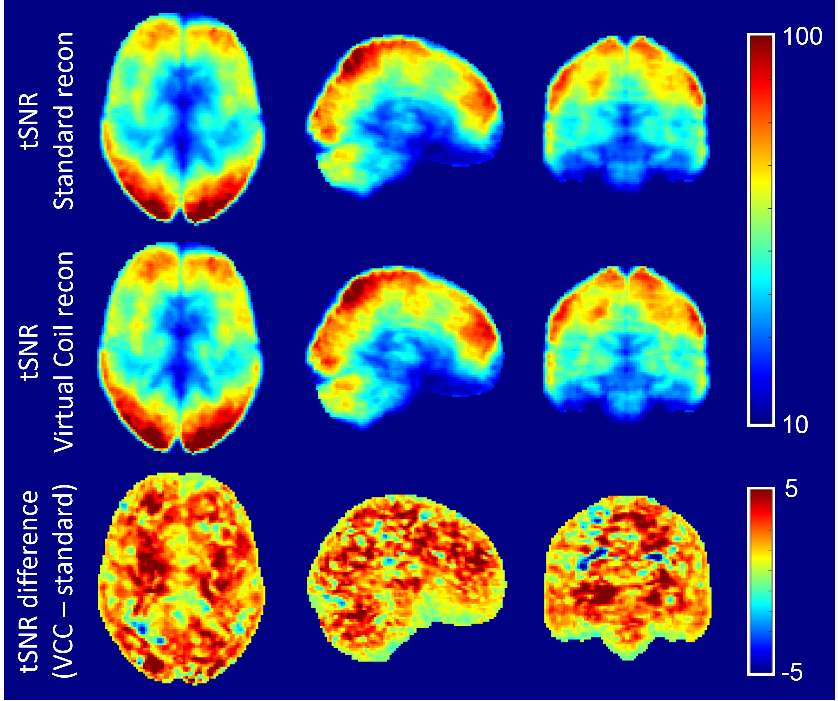
Mean tSNR maps across subjects are displayed in Figure 1 for standard and VCC reconstructions, as well as their difference. As expected from the lower g-factors of VCC reconstruction, tSNR is increased in almost every location in the VCC case, with a 3.4% average tSNR gain.
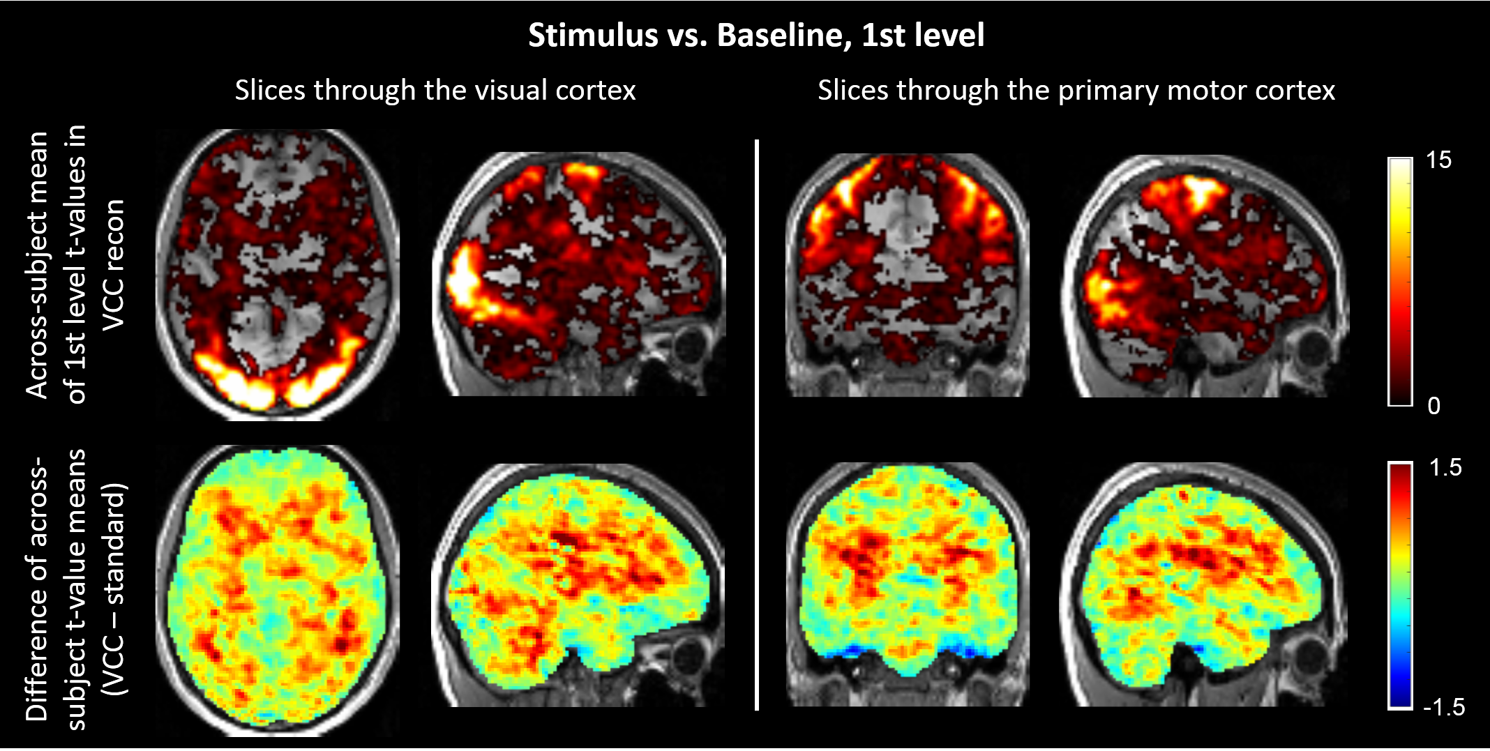
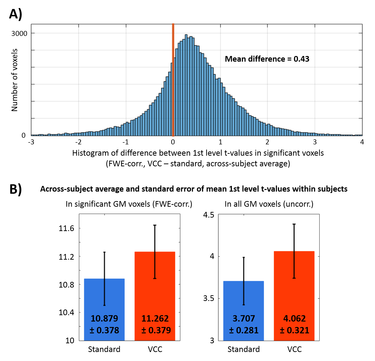
Across-subject mean of first-level t-values for stimulus vs. baseline contrast are shown in Figure 2A, using VCC reconstruction. The difference between t-values with and without using the VCC signals (Figure 2B) shows a general increase in t-values when using VCC reconstruction, especially in deep brain areas. This visual inspection is supported by Figure 3A, where the histogram of t-value differences between VCC and standard reconstructions is shifted to positive values, indicating an overall increase in t-values in the VCC case. In Figure 3B, the across-subject averages of within-subject mean t-values are plotted, pointing out the increase of mean t-values in the VCC reconstruction.
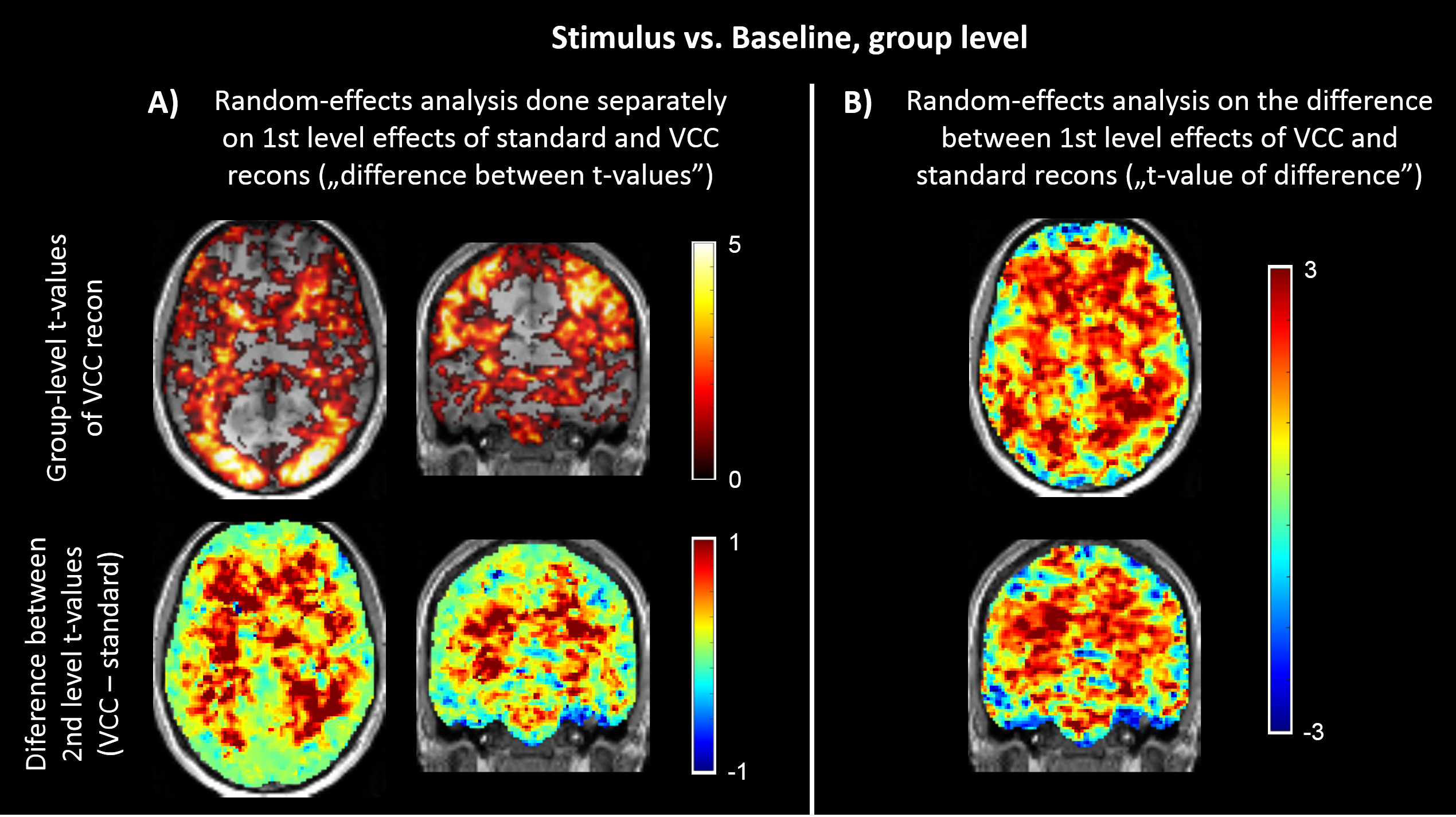
Separate group-level random-effects analyses of standard and VCC reconstructions (Figure 4A) show increased group-level t-values in deep brain areas in for VCC reconstruction, with minor improvements in the activated areas. However, the random-effects analysis performed on the differences between first-level effects of VCC and standard reconstruction (Figure 4B) indicate that effect sizes are consistently higher in VCC reconstruction in most brain regions.
Conclusion
We have shown that Virtual Conjugate Coil reconstruction can provide statistical improvement in accelerated fMRI. First- and group-level effect sizes, t-values and temporal SNR are increased using VCC reconstruction in the tested visuomotor task. Our results indicate that the benefit of VCC reconstruction could be even higher for deep brain activations, where the advantage of VCC in noise reduction is usually greater.Acknowledgements
AK, PH and PV were supported by a grant from the Hungarian Brain Research Program (KTIA_13_NAP-AI/18) to ZV. The authors thank Edward J. Auerbach, Junqian Xu, Essa Yacoub, Steen Moeller and their colleagues at Center for Magnetic Resonance Research, University of Minnesota for the blipped-CAIPI SMS EPI sequence.References
1. Setsompop K, Gagoski BA, Polimeni JR, Witzel T, Wedeen VJ, Wald LL. Blipped-controlled aliasing in parallel imaging for simultaneous multi-slice echo planar imaging with reduced g factor penalty. Magn Reson Med. 2012;67(5):1210–1224.
2. Moeller S, Yacoub E, Olman CA, Auerbach EJ, Strupp J, Harel N, Ugurbil K. Multiband multislice GE-EPI at 7 tesla, with 16-fold acceleration using partial parallel imaging with application to high spatial and temporal whole-brain fMRI. Magn Reson Med. 2010;63(5):1144-1153.
3. Feinberg DA, Moeller S, Smith SM, Auerbach EJ, Ramanna S, Glasser MF, Miller KL, Ugurbil K, Yacoub E. Multiplexed Echo Planar Imaging for Sub-Second Whole Brain FMRI and Fast Diffusion Imaging. PLoS One. 2010;5(12):e15710.
4. Xu J, Moeller S, Auerbach EJ, Strupp J, Smith SM, Feinberg DA, Yacoub E, Ugurbil K. Evaluation off slice accelerations using multiband echo planar imaging at 3 T. Neuroimage. 2013;83:991-1001.
5. Pruessmann KP, Weiger M, Scheidegger MB, Boesiger P. SENSE: sensitivity encoding for fast MRI. Magn Reson Med. 1999;42(5):952–962.
6. Blaimer M, Gutberlet M, Kellman P, Breuer FA, Köstler H, Griswold MA. Virtual coil concept for improved parallel MRI employing conjugate symmetric signals. Magn Reson Med. 2009;61(1):93-102.
7. Blaimer M, Choli M, Jakob PM, Griswold MA, Breuer FA. Multiband Phase Constrained Parallel MRI. Magn Reson Med. 2013;69(4):974-980.
8. Kettinger AO, Setsompop K, Kannengiesser SAR, Breuer FA, Vidnyanszky Z, Blaimer M. Combining Virtual Conjugate Coil reconstruction with partial Fourier imaging for maximized utilization of k-space conjugate symmetry. Proceedings of the 34th Annual Scientific Meeting of the ESMRMB, Barcelona, Spain; 2017. p.179.
9. Penny WD, Friston KJ, Ashburner, JT, Kiebel SJ, Nichols TE. Statistical parametric mapping: the analysis of functional brain images. Elsevier. 2017.
10. Todd N, Moeller S, Auerbach EJ, Yacoub E, Flandin G, Weiskopf N. Evaluation of 2D multiband EPI imaging for high-resolution, whole-brain, task-based fMRI studies at 3T: Sensitivity and slice leakage artifacts. Neuroimage. 2016;124(Pt A):32-42.
11. Auerbach EJ, Xu J, Yacoub E, Moeller S. Multi-band accelerated 2D EPI pulse sequence, version R015, released 3 March 2017, acquired through C2P contract from Center for Magnetic Resonance Research, Department of Radiology, University of Minnesota. https://www.cmrr.umn.edu/multiband/
Figures