5435
Potential role of diffusion tensor imaging in assessing neonatal neurobehavior development in the newborn stage1Department of Diagnostic Radiology, The first Affiliated Hospital of Xi'an Jiaotong University, Xi'an, China, 2Department of Biomedical Engineering, School of Life Science and Technology, Xi’an Jiaotong University, Xi'an, China
Synopsis
Maturation of brain structure underpins its functionality. From birth, brain structure and neurobehavior abilities progressively mature. However, specific link between these two entities remains unclear. This study therefore aims to investigate such correlation between neonatal neurobehavior and brain white matter (WM) microstructural development measured by diffusion tensor imaging (DTI)-fractional anisotropy (FA). Results indicated that neonatal behavior abilities significantly correlated with sensorimotor WM FAs; while active-tone just correlated with motor-associated WM. These findings demonstrate the specialized correlations of specific neurobehavior abilities with microstructural maturation in corresponding WMs, suggesting the potential role of DTI in assessing the neonatal neurodevelopment in clinical practice.
Introduction
Maturation of brain structure underpins its functionality1,2. From birth, brain structure and neurobehavior performance progressively develop and get matured1. However, synergistic link between these two entities especially in the early neonatal period remains unclear. Previous studies have concluded the maturation progression of brain white matter (WM), i.e. fiber growth, pruning and myelination1. In the newborn stage (the first postnatal month), fiber pruning and myelination occur in most WMs. However, whether the developing WMs synchronously get functionalization and present specialized neurobehavior abilities remains unknown. To address this issue, specific correlations between neonatal neurobehavior performances (two items: behavior and active tone) and brain WM microstructural development as measured by diffusion tensor imaging (DTI)-derived fractional anisotropy (FA), are investigated in this study.Methods
The Institutional Review Board approved this study and all the written informed consents were obtained from parents of neonates. Populations One hundred and one neonates with no abnormality on MRI were included. MR Protocols All MR imaging examinations were performed using a 3.0T scanner (Signa HDxt, GE Healthcare, Milwaukee, Wisconsin) with an 8-channel head coil. The protocols included sequences: (1) a transverse 3D T1-weighted sequence (TR/TE= 10 ms/4.6 ms, matrix= 256×256, section-thickness= 1 mm, FOV= 240 mm); (2) a 2D T2-weighted sequence (TR/TE= 4200 ms/120 ms; matrix= 256×256; section-thickness= 4 mm; FOV= 180 mm); (3) Diffusion tensor imaging (DTI protocol1: 35 directions, b-value= 1000 s/mm2, TR/TE= 5500 ms/95 ms, section-thickness= 4 mm, FOV= 180 mm, matrix= 128×128; DTI protocol2: 38 directions, b-value= 600 s/mm2, TR/TE = 11000 ms/69.5 ms, section-thickness = 2.5 mm, FOV= 180 mm, matrix= 128×128). Data and statistical analysis DTI data was processed with the aid of the FMRIB software library (FSL, www.fmrib.ox.au.uk/fsl). Combat harmonization4 for DTI-FA between two DTI protocols were performed because of their significant differences (P<0.05).The tract-based spatial statistics (TBSS) was used to investigate the relationships between FA and neurobehavioral scores (i.e. behavioral and active)4,5. Spearman correlation was further used to explore the relationship between neurobehavioral scores and FA along the typical visual, auditory and sensorimotor WMs, i.e. OR (optical radiation), AR (auditory radiation), CST (corticospinal tract) and thal-PSC (thalamus-primary somatosensory cortex). These four tracts were extracted by automated fiber quantification (AFQ).All statistical analysis were performed by using using MATLAB software (version R2012b; Mathworks, Inc.; Natick, MA). P<0.05 was considered statistically significant.Results
One hundred and one neonates (male/female, 28/22; GA range, 30~42weeks) with no abnormalities on MRI within 28 days after birth were included (Table 1).
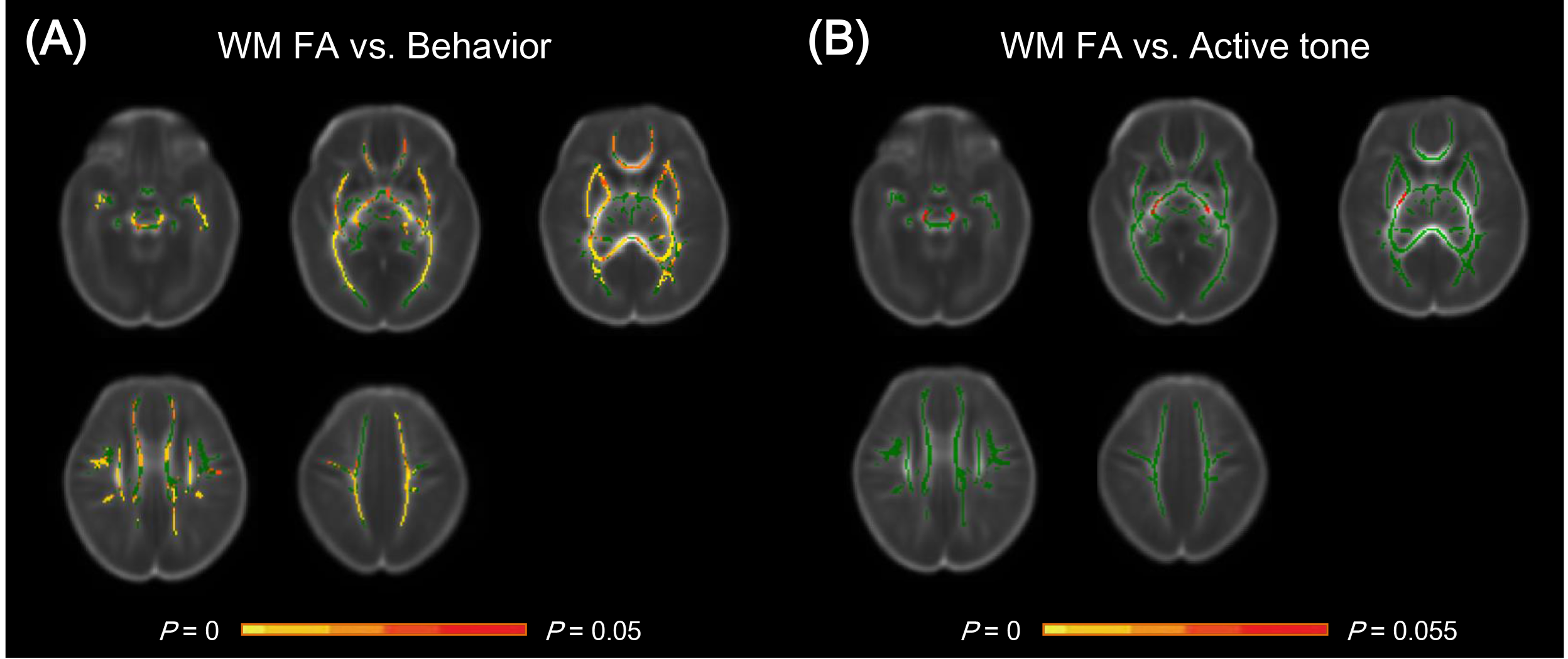
Using TBSS, behavior scores (score range, 6-12) of neonates showed significant correlations with FA in regional WM, e.g. corpus callosum, corticospinal tract, optical radiation, auditory radiation and etc. (P<0.05); while, boundary significant correlations were observed for active tone (score range, 2-8) in local WM regions, i.e. cerebral peduncle and posterior limb of internal capsule (P=0.055). (Figure 1)
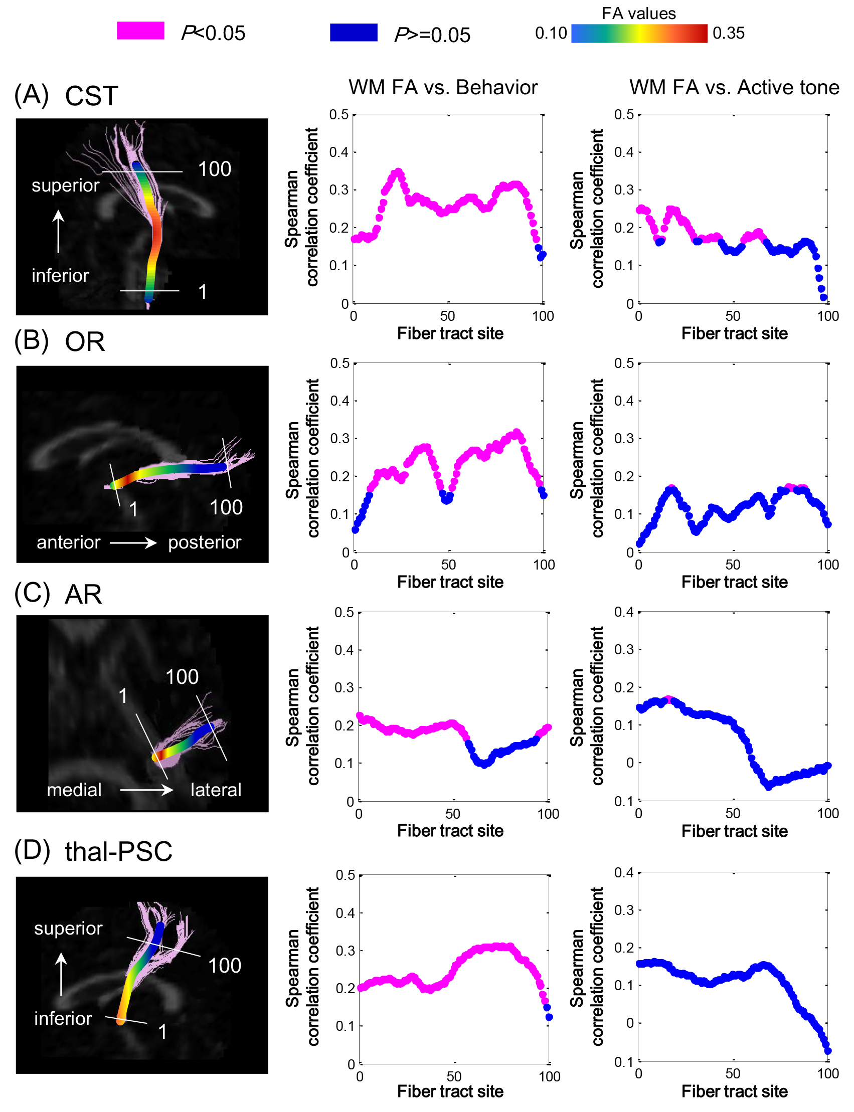
AFQ results indicated that FA along the almost entire CST, OR and thal-PSC showed significant correlations with behavior scores (CST: r_mean = 0.26 [range, 0.17-0.35]; OR: r_mean = 0.24 [range, 0.17-0.32]; thal-PSC: r_mean = 0.25 [range, 0.17-0.31]; P<0.05); while significant correlations mainly located in the initial and middle segments of AR (r_mean = 0.19 [range, 0.17-0.22]; P<0.05). For active tone, significant correlations were only observed in the initial and middle segments of CST (r_mean = 0.20 [0.17-0.25]; P<0.05). (Figure 2)
Discussion
This study indicated that neonatal behavior performance presents significant correlation with the maturation of the sensorimotor WMs, i.e. the CST, OR, AR and thal-PSC; while active tone just correlates with motor-associated CST. Brain sensorimotor WMs launch the myelination and progressively get maturation in the newborn stage1. In the items of behavior assessment, neonatal visual, auditory, sensory and motor responses have all been evaluated3,4. Significant correlations between sensorimotor WM FA and behavior assessment indeed demonstrated the functional maturation of sensorimotor WMs. For the active tone reflecting the motor ability3,4, significant relationship with CST FA was found. This further suggested the specialization of the motor-associated brain structure-function.
Notably, AR shows significant correlation with behavior at the initial and middle sections of fiber tracts; while almost the whole sections were included in others. These may be rooted in the sequence and lasting time of myelination1. Specifically, AR initials the myelination from central/proximal portion to peripheral/distal one; and presents a longer myelination than CST and OR. It may be these facts that led to no correlation with behavior assessment in the terminal part of AR.
Conclusion
Our findings demonstrate the specialized correlations of specific neurobehavior abilities with microstructural maturation in corresponding WMs, suggesting the potential role of DTI in assessing the neonatal neurodevelopment in clinical practice.Acknowledgements
This work was supported by the National Key Research and Development Program of China (2016YFC0100300), National Natural Science Foundation of China (No. 81471631, 81771810 and 51706178), China Postdoctoral Science Foundation (No. 2017M613145), and Shaanxi Provincial Natural Science Foundation for Youths of China (No. 2017JQ8005).References
- Dubois J, Dehaene-Lambertz G, Kulikova S, et al. The early development of brain white matter: a review of imaging studies in fetuses, newborns and infants. Neuroscience, 2014, 276: 48-71.
- Qiu A, Mori S, Miller MI. Diffusion tensor imaging for understanding brain development in early life. Annu Rev Psychol, 2015, 66: 853-876.
- Fortin JP, Parker D, Tunç B, et al. Harmonization of multi-site diffusion tensor imaging data. Neuroimage 2017;161:149-170.
- Bao XL, Yu RJ, Li ZS, Zhang BL. Twenty-item behavioral neurological assessment for normal newborns in 12 cities of China. Chin Med J, 1991, 104:742-746.
- Dubowitz LMS, Dubowitz V, Mercuri E. The neurological assessment of the preterm and full-term newborn infant. 2nd ed. London: Mac Keith Press, 1999.
Figures