5433
Establishing a developmentally appropriate fMRI paradigm for presurgical mapping of memory in childrenAmanda G Wood1, Elaine Foley2, Parnpreet Virk2, Helen Ruddock3, Paras Joshee4, Kelly Murphy2, and Stefano Seri2
1Life and Health Sciences, Aston Brain Centre, Aston University, Birmingham, United Kingdom, 2Aston University, Birmingham, United Kingdom, 3University of Liverpool, Liverpool, United Kingdom, 4University of Birmingham, Birmingham, United Kingdom
Synopsis
The use of functional MRI to evaluate risk to memory function following temporal lobe surgery in children cannot rely on adult tools. We present a novel fMRI paradigm that is brief, independent of reading ability, and therefore a candidate for presurgical evaluation. Data from 36 adults and 19 children (all healthy controls) show that the paradigm captures the expected leftward asymmetry of mesial temporal activation in adults. A more symmetrical pattern is observed in children, consistent with the emergence of hemispheric specialisation across childhood. These data have important implications for the interpretation of presurgical memory fMRI in the paediatric setting.
Introduction
Functional magnetic resonance imaging (fMRI) is used increasingly in planning for temporal lobe surgery, including memory paradigms to evaluate the impact of resection on post-operative memory functions. The use of fMRI paradigms to elicit activation in eloquent cortex is complicated by the expected lower general cognitive skills of children, the impairments exhibited in those with temporal lobe epilepsy (TLE) and the normal representation of function during development, which may differ from adults. Data from adults with epilepsy suggest that fMRI can aid prediction of postoperative memory, however existing paradigms are not suitable for children. We aimed to evaluate the pattern of activation using a novel developmentally-appropriate memory paradigm. In doing so, we also aimed to determine whether the lateral representation of verbal memory activation differed in young children compared to older participants. We predicted that young children would exhibit bilateral mesial temporal activation during the task compared to adults, who would show typical asymmetric activation.Methods
Participants were adults and children without a history of neurological diagnosis recruited through community advertisements. Adults and parents/guardians of minors provided written informed consent and all children assented to the study. Data were acquired at two centres, Birmingham University Imaging Centre (BUIC; Philips Achieva 3 t MRI) and Aston Brain Centre (ABC; Siemens Trio Tim, 3 t system), UK. Whole brain, high-resolution T1-weighted images were acquired for image co-registration. Functional MRI data were acquired with an echo-planar acquisition protocol to measure the blood-oxygen level dependent response (ABC: TR 4110, delay 2300; TE 30; 2 mm isotropic voxels; 26 slices (run length ~3 minutes 9 seconds). BUIC: TR 4000, delay 2300; TE 35, 2.3*2.3*2.5mm isotropic voxels; 26 slices). The fMRI paradigm comprised an incidental verbal encoding task presented aurally. The overall study design involved three phases: 1) Out of scanner familiarisation, 2) In-scanner encoding, 3) out of scanner recognition. Words were selected on the basis that they contained 1 to 2 syllables and had an age of acquisition (AoA) rating less than 600. Half of the words selected were ‘living’ and half were ‘non-living’ and these were equated for AoA, familiarity, imagibility, and length. During familiarisation, participants listened to two words presented on a laptop via headphones. They pressed a button to indicate whether words were living or non-living to ensure that rehearsal strategies were not employed differentially in adults and children and encourage deep encoding via semantic access. This established them as the ‘familiar’ words for the subsequent ‘novel’ versus ‘familiar’ contrast. During scanning, words were presented in alternating blocks of ‘novel’ and ‘familiar’ (ie repeated) items, with participants responding ‘living’ or ‘non-living’ via button-press. To overcome scanner noise, words were presented during the TR delay. Following scanning, a surprise post-recognition memory test (‘old’ fMRI words versus ‘new’ foils) determined which items were successfully encoded, for later analysis. Only correctly recognised words were included in further analyses. Data were analysed with SPM12 using the General Linear Model. The key contrast was between encoded novel versus familiar words. Group-level analyses controlled for scanner site. Lateralisation indices were estimated using the SPM LI Toolbox using temporal and hippocampal masks (derived from Marsbar). The relationship between child age and asymmetry of activation was then evaluated by calculating LI scores for individual cases at the threshold at which the average LI value was determined in the bootstrapped method.Results
Data from 36 right-handed adults and 19 children (aged 8-15 years) are presented. Adults showed the expected pattern of leftward activation within the mesial temporal lobe (p<0.001 corrected, T=3.32; Figure 1). Their average bootstrapped LI score for the temporal mask was 0.65 and hippocampal mask 0.88. By contrast, children showed a more bilateral pattern of activation within the mesial temporal lobe (Figure 2), with an average bootstrapped LI score for the temporal mask of 0.3 and 0.49 for the hippocampal mask. There was a positive but non-significant association between children’s age and hippocampal LI score (r=0.504, p=0.06).Discussion
This study highlights the importance of establishing child-friendly tasks that can be used in future clinical studies. The paradigm is brief and yet provides reliable activation in adults and children. The more symmetric activation in children reflects a co-occurrence of increased leftward representation of verbal functions across typical development. This implies that bilateral activation in young children cannot be interpreted as functional reserve of the contralateral temporal lobe, which has significant implications for clinical management of young children requiring temporal lobe resection. Future work to establish the prognostic value of the paradigm in paediatric surgical candidates is required.Acknowledgements
Professors Wood and Seri and Dr Foley wish to acknowledge funding support from Epilepsy Research UK (pilot program).References
No reference found.Figures
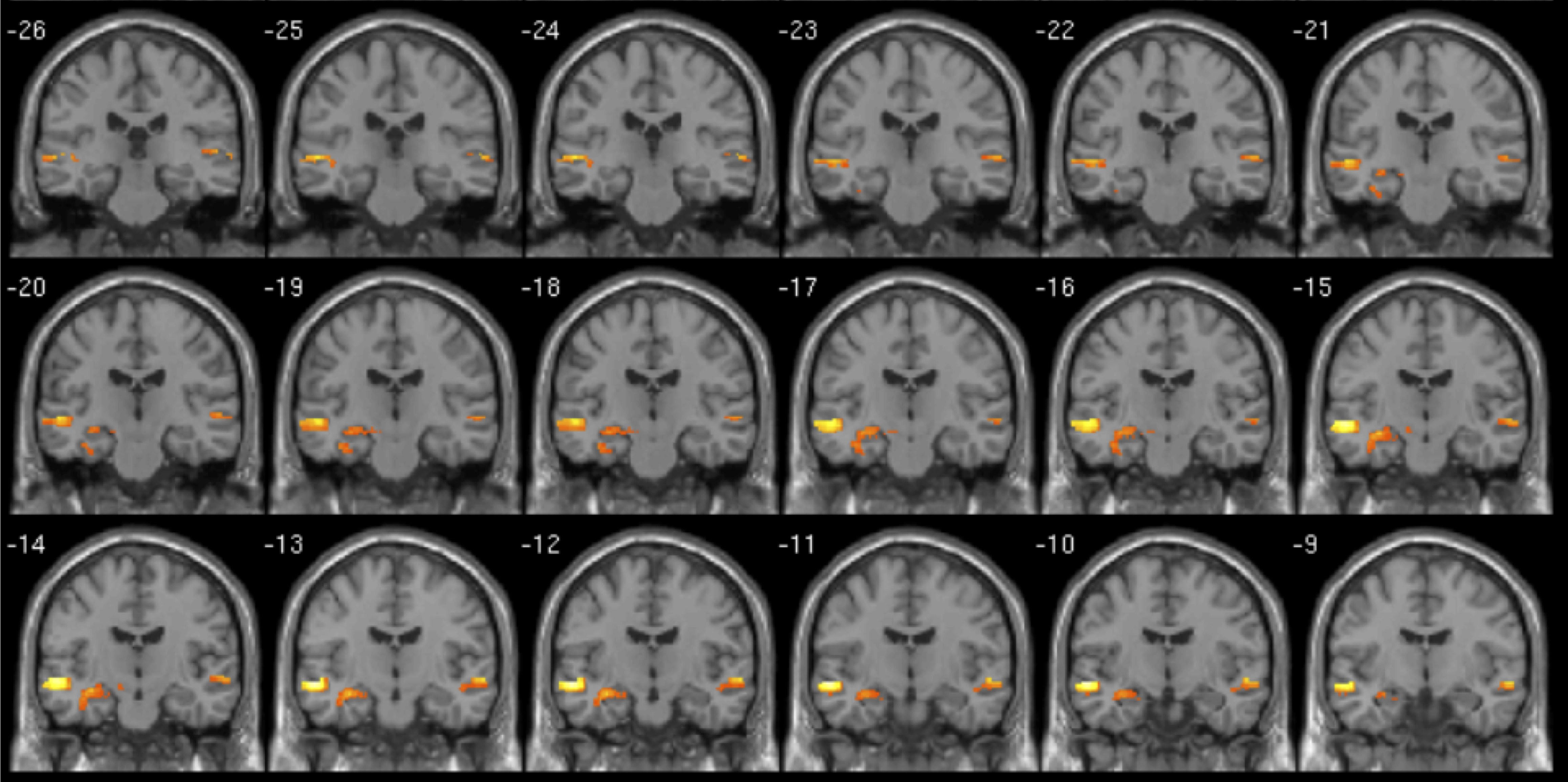
Activation in 36 right-handed adults during verbal encoding. Novel>familiar contrast (p<0.001, corrected) for correctly recognised words. L=L
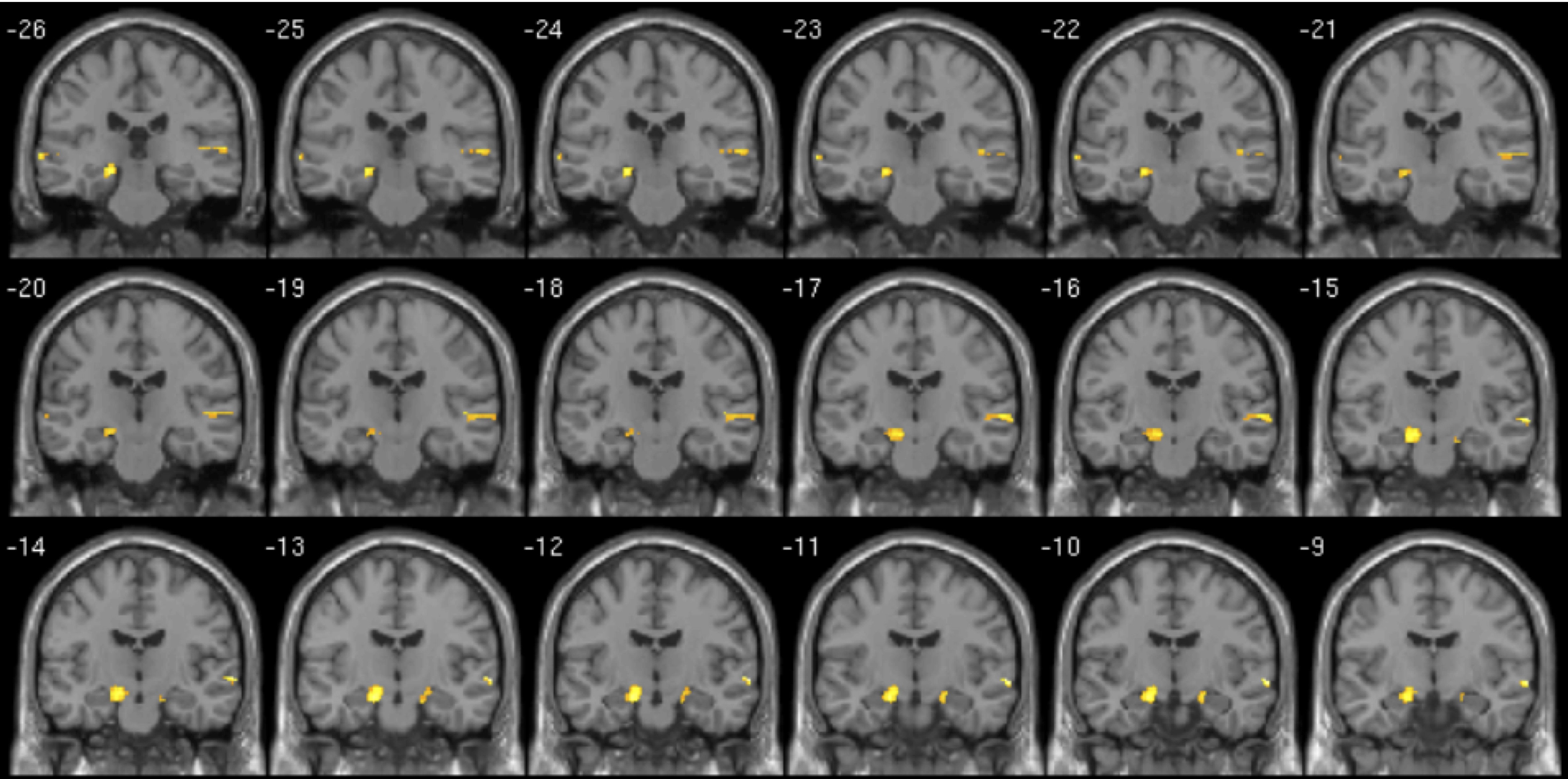
Activation in 19 right-handed children (aged 8-15 years) during verbal encoding. Novel>familiar contrast (p<0.001, corrected) for correctly recognised words. L=L