5431
Differential effects of hunger and depression on cerebral blood flow in healthy adolescents1Unité de recherche sur les comportements et mouvements anormaux (URCMA, IGF, INSERM U661 UMR 5203), Departments of neurosurgery, Montpellier University Hospital Center, Gui de Chauliac Hospital, University of Montpellier, Montpellier, France, 2Institut d’Imagerie Fonctionnelle Humaine, I2FH, Department of Neuroradiology, Montpellier University Hospital Center, Gui de Chauliac Hospital, University of Montpellier, Montpellier, France, 3Unité de pathologie cérébrale résistante, Department of neurosurgery, Montpellier University Hospital Center, Montpellier, France, 4Siemens Healthcare GmbH, Application Development, Erlangen, Germany
Synopsis
This study aims to explore the appetite effect on taste and depression on healthy adolescents using Arterial Spin Labeling. Fifteen participants complete the Multiscore Depression Inventory for Children test and two MRI sessions: pre-lunch (hunger) and post-lunch (satiety). We found an increased CBF – cerebral blood flow – during hunger in the posterior insula (anticipation and motivation of feeding) and during satiation in the precuneus, lingual gyrus and cuneus (inhibition pattern of food intake). We show that the correlations between depression and CBF are modulated by appetite in the precuneus, operculum, lingual, cuneus, middle frontal gyrus and inferior parietal lobule.
Introduction
Obesity, defined as Body Mass Index>30kg/m², is a major public health concern which can be linked to perturbed eating behavior1. However, our knowledge is rather limited and it is first necessary to investigate the basis of appetite in healthy population especially in children and adolescents. It has been shown that hunger leads to an increased CBF – cerebral blood flow – in the hypothalamus, thalamus, putamen, insular cortex, orbitofrontal cortex and limbic areas2,3. Satiation is associated with a greater CBF in the prefrontal cortex and inferior parietal lobule. In addition, it is crucial to remind that the CBF could be modified by psychological aspects especially, depression. No study questioned this aspect in the context of feeding.
Therefore, the aim is to extend knowledge of the cognitive processes involved in the food intake in healthy adolescents using Arterial Spin Labeling (ASL). We expect that the cerebral blood flow in the taste areas: (i) is implicated in hunger states compared to satiety states and (ii) is influenced by depression.
Methods
Fifteen healthy right-handed adolescents (7 female; 8 male; age=14.1±1.3 years, mean±SD) undergo two MRI sessions: (i) a pre-lunch called “hunger” condition and (ii) post-lunch called “satiety” condition. They are restricted from eating for 4h or 5h before the first MRI session, and then a standardized lunch is given at 1:00 pm. Two hours later, the second MRI session is performed. Neuroimaging data are collected on a 3T magnet (MAGNETOM Skyra, Siemens, Healthcare, Germany) with a 32 channels head coil. Before MRI, depression is assessed using the Multiscore Depression Inventory for Children including the anxiety, self-esteem, sad mood, instrumental helplessness, social introversion, low energy, pessimism and defiance subscales.
3D pseudo continuous ASL was performed using a prototype sequence to estimate CBF offline through a standard processing pipeline5. Correction is performed for white matter lesions and partial volume effect. Required tissues segmentations are obtained from the high resolution 3DT1 using the SPM12 software. The normalized CBF maps are entered into the whole brain voxel-wise general linear modeling analysis which included paired-t-tests and voxels based morphometry analysis.
Results
During the “hunger” condition relative to the “satiety” condition, the CBF is significantly increased in bilateral posterior insula (figure 1&2). Conversely, compared to the “hunger” condition, the “satiety” condition shows a significantly stronger CBF within the right cuneus, left lingual gyrus and left precuneus.
Correlations with depression scores are observed during the “hunger” condition (figure 3). Concerning the total depression scores, a positive correlation with CBF is found in the bilateral precuneus, left cuneus, right operculum gyrus and right posterior cingulate gyrus. For the low energy scores, a positive correlation with CBF is identified in the left superior temporal gyrus, left middle frontal gyrus, left operculum gyrus, left anterior insula and right posterior insula. For the self-esteem scores, a positive correlation with CBF is observed in the bilateral operculum gyrus. Regarding the sad mood scores, a negative correlation with CBF is found in the left middle frontal gyrus. Furthermore, we report only one correlation during the “satiety” condition. A negative correlation between self-esteem scores and CBF is observed in the left middle frontal gyrus and left inferior parietal lobule.
Discussion
Results revealed that hunger is associated with increased CBF in the bilateral posterior insula, areas previously described as important in the anticipation and motivation of feeding3. In contrast, satiation is associated with increased CBF in the left precuneus, left lingual gyrus and right cuneus (inhibition pattern of food intake2). Furthermore, we show that the correlations between depression scores and CBF are modulated by appetite in the bilateral precuneus, bilateral operculum, left lingual, left cuneus, left middle frontal gyrus and left inferior parietal lobule. In concordance with the literature6,7, our findings suggest that participants with a higher depression scores showed greater incentive and anticipation to eat, higher irritability levels, and required more mental effort to endure hunger.Conclusion
To conclude, our work confirms the involvement of posterior insula and precuneus in the modulation of appetite. We also highlight an unpublished finding that the feeling of hunger could modulate the relation between depression and brain activity in specific regions as precuneus, operculum, lingual, cuneus, frontal and parietal regions. It would be interesting to perform a functional connectivity analysis between these regions, but also taking into account the CBF modifications related to the hunger and psychological aspects like depression.Acknowledgements
No acknowledgement found.References
1. Ogden, C. L. et al. Prevalence of Overweight and Obesity in the United States, 1999-2004. JAMA 295, 1549–1555 (2006).
2. Tataranni, P. A. et al. Neuroanatomical correlates of hunger and satiation in humans using positron emission tomography. Proc. Natl. Acad. Sci. U. S. A. 96, 4569–4574 (1999).
3. Del Parigi, A. et al. Neuroimaging and obesity: mapping the brain responses to hunger and satiation in humans using positron emission tomography. Ann. N. Y. Acad. Sci. 967, 389–397 (2002).
4. Monkul, E. S. et al. Abnormal Resting State Corticolimbic Blood Flow in Depressed Unmedicated Patients With Major Depression: A 15O-H2O PET Study. Hum. Brain Mapp. 33, 272–279 (2012).
5. Asllani, I., Borogovac, A. & Brown, T. R. Regression algorithm correcting for partial volume effects in arterial spin labeling MRI. Magn. Reson. Med. 60, 1362–1371 (2008).
6. Porubská, K., Veit, R., Preissl, H., Fritsche, A. & Birbaumer, N. Subjective feeling of appetite modulates brain activity: an fMRI study. NeuroImage 32, 1273–1280 (2006).
7. Besteher, B. et al. Brain structural correlates of irritability: Findings in a large healthy cohort. Hum. Brain Mapp. (2017). doi:10.1002/hbm.23824
Figures
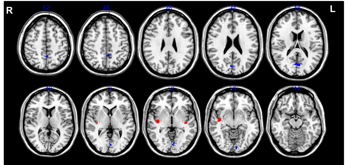
Figure 1: Statistical parametric maps showing regions of increased CBF in the “hunger” condition and “satiety” condition.
During “hunger” condition relative to “satiety” condition, CBF increases in the bilateral posterior insula (red). During the “satiety” condition relative to the “hunger” condition, CBF increases in the right cuneus, left lingual gyrus and left precuneus (blue). Statistical threshold is used for a p-value p<0.0005 and a minimum cluster size of 30 voxels, uncorrected. Results are adjusted for age and sex.
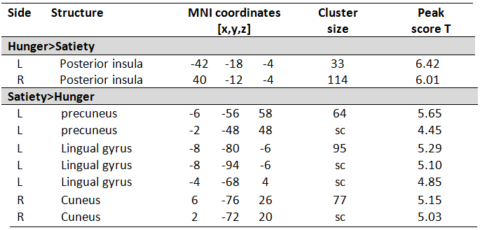
Figure 2: Brain regions showing an increased CBF in the “hunger” condition and “satiety” condition.
Results are assessed using paired t-test analyses by adjusting for age and sex. Statistical threshold is used for a p-value p<0.0005 and a minimum cluster size of 30 voxels, uncorrected. Results are adjusted for age and sex. Abbreviation: sc, same cluster.
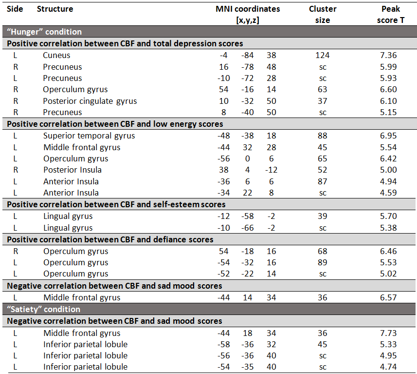
Figure 3: Brain regions showing a correlation between brain perfusion and depression scores for “hunger” and “satiety” conditions.
Correlations are reported between cerebral blood flow and (a) total depression score, (b) low energy, (c) self-esteem, (d) defiance and (e) sad mood for each condition. No correlation is found with the anxiety, instrumental helplessness, social introversion and pessimism. Results are assessed using voxel based morphometry analyses by adjusting for age and sex. Statistical threshold is used for a p-value p<0.0005, k=30 voxels, uncorrected. Abbreviation: sc, same cluster.