5424
Chemotherapy reduces microstructural asymmetry in the brain1Diagnostic Imaging, St Jude Children's Research Hospital, Memphis, TN, United States, 2Biostatistics, St Jude Children's Research Hospital, Memphis, TN, United States
Synopsis
Previously, we discovered two types of brain microstructural asymmetry: myelin-related asymmetry and axon-related asymmetry in healthy volunteers. In this study, we found that chemotherapy with methotrexate significantly reduced the microstructural asymmetries in patients with acute lymphoblastic leukemia, having a greater impact on asymmetries in younger patients than in older ones. Our results indicate that chemotherapy may lead to atypical brain development long before patients become symptomatic. These results may help to provide insights into neurocognitive deficits caused by chemotherapy.
Introduction
Brain microstructural asymmetry can be measured using DTI. Previously, we discovered two types of brain microstructural asymmetry: myelin-related asymmetry and axon-related asymmetry in healthy volunteers. Axon asymmetry exists in both the front and back brain, whereas myelin asymmetry is more pronounced in the back brain. Chemotherapy preferentially damages the white matter of the brain1,2. In this study, we showed that chemotherapy with methotrexate substantially degraded the axon and myelin asymmetry in the patients with acute lymphoblastic leukemia (ALL) in comparison with healthy volunteers.Methods
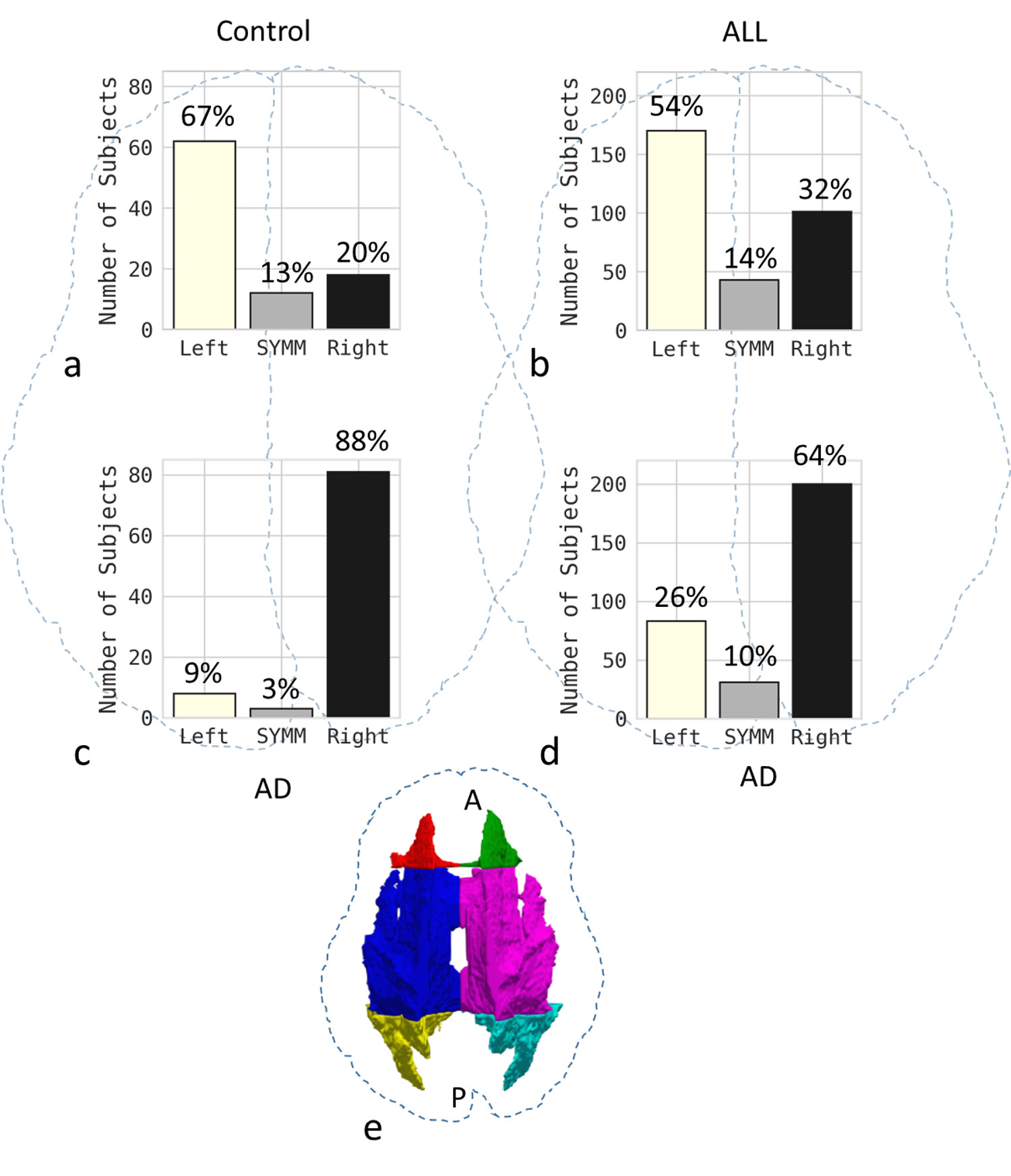
We evaluated microstructural asymmetry in a total of 314 patients with ALL (190 male and 124 female subjects aged 1 to 19 years) and 92 controls (54 male and 38 female subjects aged 6 to 25 years). DTI was performed using a SE-EPI sequence with 12 directions. The DTI protocol parameters were as follows: TR = 6500 ms; TE = 120 ms; b = 0, 700 s/mm2; 3-mm slice thickness; 40 axial slices; in-plane resolution of 1.5 × 1.5 × 3.0 mm. T1w images were acquired with an in-plane resolution of 0.82 × 0.82 mm. DTI images were first registered to T1w images then further registered to the MNI 152 atlas using FSL. The white matter was parcellated into three regions: front, middle, and back as shown in Fig. 1e3.
The asymmetry index (AI) parameter was calculated using $$$AI=(P_L-P_R)/C$$$ . $$$P_L$$$ is the averaged DTI parameter on the left side, $$$P_R$$$ is the averaged DTI parameter on the right side, and $$$C$$$ is the normalization constant, which is equal to 1.0 × 10⁻³ mm2/s for Axial diffusivity (AD), MD, and radial diffusivity (RD), and to 1.0 for FA.
Results
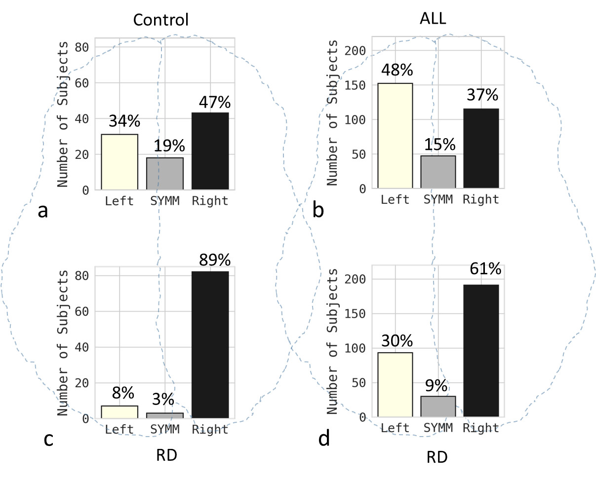
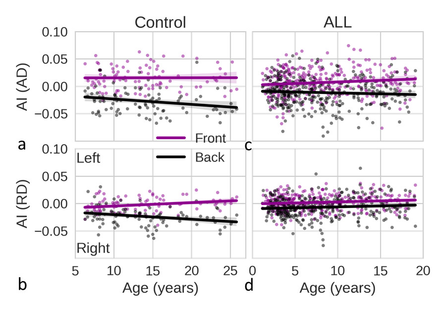
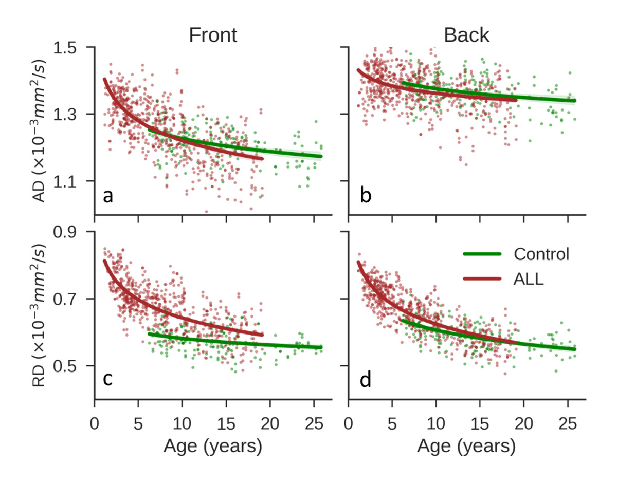
AD asymmetry is referred to as axon asymmetry and RD asymmetry is referred to as myelin asymmetry since AD and RD differentiate axonal and myelin injuries, respectively4. Fig. 1 shows that microstructural axon asymmetries in ALL patients were decreased in both front and back brain in comparison with control group. The differences in percentage between two sides were 22% for ALL and 57% for control in the front brain, 38% for ALL and 79% for control in the back brain. Fig 2 shows that microstructural myelin asymmetries in the back brain in ALL patients were also decreased in comparison with control group. Fig 3 (a, c) shows that the absolute asymmetry index of AD in ALL patients were smaller than that in controls for the whole range of age. Fig 3 (b, d) shows that the absolute asymmetry index of RD in the back brain of ALL patients were smaller than that in controls. In Fig 4, the RD values of subjects in the ALL cohort were significantly larger (P < 0.01) than those of subjects in the control cohort, indicating that RD was more sensitive to the brain damage and that chemotherapy might cause more damage to myelin than to axons. On comparing the two cohorts, the difference in RD in the front brain was much larger than the difference in the back brain (compare Fig. 4c and 4d), which might indicate that there was greater myelin damage in the front brain. Finally, the RD in younger patients with ALL differed from that in controls of the same age to a greater extent than did the RD in older patients with ALL (Fig. 4c). These differences decreased with increasing age, indicating that chemotherapy may cause more severe damage to younger patients than to older ones.Discussion / Conclusion
We have shown that chemotherapy can greatly reduce microstructural asymmetry in patients with ALL, as compared with healthy controls. This is the first report of changes in brain structural asymmetry that are related to chemotherapy in patients with ALL. ALL survivors diagnosed at a younger age are at a larger risk for neurocognitive deficits than those diagnosed at older age5. This is consistent with our observation that the younger patients in our study had more myelin damage than did the older patients when compared to controls. All these findings support the development of personalized treatment and early intervention, especially for younger patients, to reduce the incidence and severity of cognitive impairment.Acknowledgements
We thank Keith A. Laycock for editorial assistance. This work was supported by RO1 CA90246 and Cancer Center Support Grant P30 CA-21765 from the National Cancer Institute, National institutes of Health, Bethesda, MD, and by the American Lebanese Syrian Associated Charities (ALSAC), Memphis, TN.References
1. Meyers, C. A. How chemotherapy damages the central nervous system. J. Biol. 7, 11 (2008).
2. Bhojwani, D. et al. Methotrexate-induced neurotoxicity and leukoencephalopathy in childhood acute lymphoblastic leukemia. J. Clin. Oncol. 32, 949–959 (2014).
3. Reiss, A. L., Abrams, M. T., Singer, H. S., Ross, J. L. & Denckla, M. B. Brain development, gender and IQ in children. Brain 119, 1763–1774 (1996).
4. Zhang, J., Aggarwal, M. & Mori, S. Structural insights into the rodent CNS via diffusion tensor imaging. Trends Neurosci. 35, 412–421 (2012).
5. Hardy, K. K. et al. Neurocognitive functioning of children treated for high-risk B-acute lymphoblastic leukemia randomly assigned to different methotrexate and corticosteroid treatment strategies: a report from the Children's Oncology Group. J. Clin. Oncol. 35, 2700–2707 (2017).
Figures