5419
Brain growth over the first 13 years of life in typically developing and very preterm children1Victorian Infant Brain Studies (VIBeS), Murdoch Children's Research Institute, Melbourne, Australia, 2Developmental Imaging, Murdoch Children's Research Institute, Melbourne, Australia, 3Department of Paediatrics, The University of Melbourne, Melbourne, Australia, 4Florey Institute of Neuroscience and Mental Health, Melbourne, Australia, 5Department of Pediatric Newborn Medicine, Brigham and Women’s Hospital, Harvard Medical School, Boston, MA, United States, 6Clinical Epidemiology & Biostatistics Unit, Murdoch Children's Research Institute, Melbourne, Australia, 7Neonatal medicine, The Royal Children's Hospital, Melbourne, Australia, 8Department of Obstetrics and Gynaecology, The University of Melbourne, Melbourne, Australia, 9Royal Women's Hospital, Melbourne, Australia, 10Monash Institute of Cognitive and Clinical Neurosciences, Monash University, Melbourne, Australia
Synopsis
Few longitudinal cohort studies exist characterizing regional brain volumes from birth to adolescence. This study derives brain volumes in approximately 100 regions at term-equivalent, 7 and 13 years of age for 102 very preterm and 20 full-term children. The trajectory of brain development in many regions differed between very preterm and full-term children over the first 7 years of life. From 7 to 13 years brain growth slowed, ceased or regressed in both groups in a region-specific manner, apart from subcortical regions that continued to increase in volume. This study provides novel insights in typical and atypical regional brain volumetry.
Introduction
Brain growth is rapid over the first years of life and continues into adolescence1. Infants born very preterm (VP) represent a population able to provide unique insight into brain development, as preterm birth may delay or disrupt brain development2. Considering brain development occurs in a regionally-specific pattern3, it is valuable to determine how VP birth affects brain regional development.
Very few studies have followed the same group of infants from infancy to early adolescence. Our unique longitudinal cohort has tracked healthy term-born and VP infants from term-equivalent to 13 years of age. We have recently developed the Melbourne Children’s Regional Infant Brain (M-CRIB) atlas4, enabling us to accurately derive infant brain volumes that match those commonly obtained for older children and adults using the Desikan-Killiany atlas5 in FreeSurfer. Thus, we can now track growth of equivalent brain regions in infants through to adolescence.
The aim of this study was to describe the trajectory of regional brain growth in full-term and VP children from term-equivalent through to 13 years of age, and determine whether these trajectories differed between groups.
Methods
One hundred and two infants born VP (<30 weeks’ gestation) or very low birthweight (<1250 g) and 20 full-term control infants born >37 to ≤ 41 weeks’ gestation had usable MRI data at term-equivalent, 7 years, and 13 years of age corrected for prematurity.
At term (38–42 weeks' gestation), structural T2-weighted images (1.7-3.0 mm coronal slices; TR/TEs: 4000/60, 160ms) were acquired with a 1.5T MRI scanner. At 7 years, T1 images (0.85 mm sagittal slices, TR/TE: 1900/2.27ms) were obtained using a 3T MRI machine, and similarly at 13 years (0.9 mm3 sagittal slices, TR/TEs: 2530/1.77, 3.51, 5.32, 7.2ms).
Term age images were bias-corrected using N4ITK6 and brain extracted using BET7. For labelling, the M-CRIB atlas4 images were registered to each T2 image using ANTS8. PSTAPLE9 was used to apply the M-CRIB labels to each image. At 7 and 13 years of age, T1 images underwent FreeSurfer 6.0 automated segmentation5.
For each brain region, 2-level piecewise linear mixed effects regression models were used to examine the growth developmental trajectory. Group (VP or full-term) was included as a fixed effect with a random intercept to allow for the repeated observations within participants. Two different effects of time were included as continuous variables of age (years) at assessment, one before and one after 7 years. Interactions between group and the 2 time effects were included to investigate differences in trajectories between groups. Analyses were adjusted for sex. Multiple comparisons were corrected using the false discovery rate.
Results
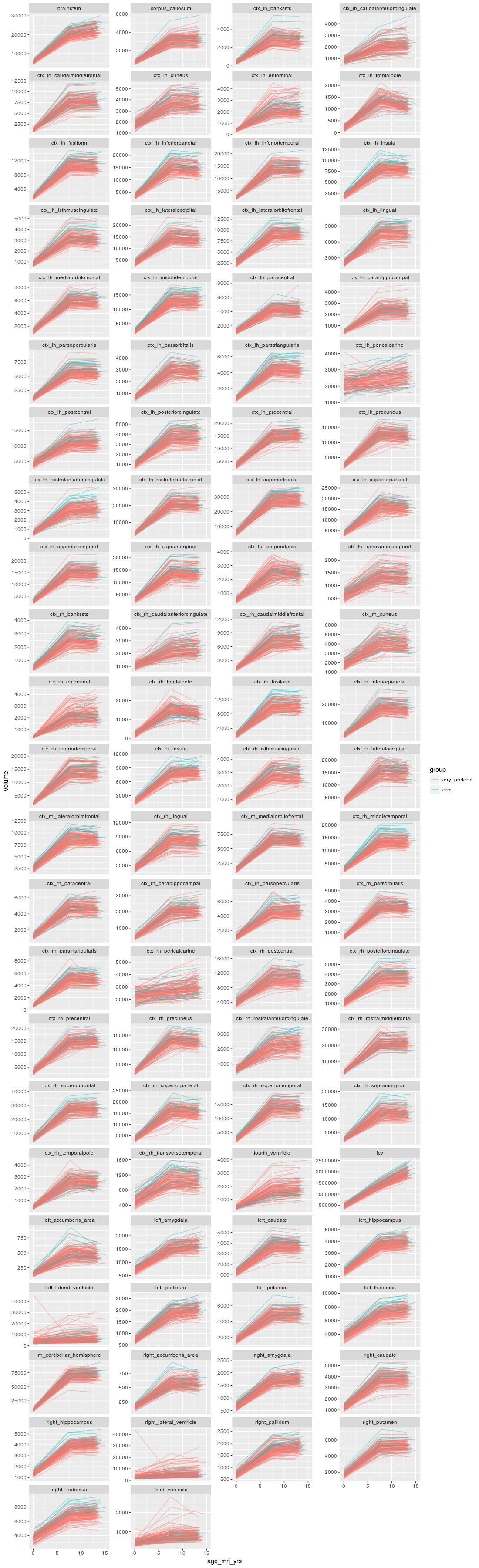
Sample characteristics are summarized in Table 1. All brain regions (excluding cerebrospinal fluid) increased in volume between term and 7 years of age (Fig. 1). There was evidence that the growth varied between groups, with faster growth in the full-term group, including basal ganglia and thalamic regions, hippocampi and corpus callosum and some frontal, parietal and temporal cortical regions. Between 7 and 13 years of age, regional volumes were relatively stable, with evidence that basal ganglia, thalamus, corpus callosum, brainstem and a few cortical regions continued to grow, while other cortical regions reduced (Fig. 1). There was little evidence that differences in volume from 7 to 13 years differed between VP and full-term groups.Discussion
The increase in brain volumes from term to 7 years of age is expected, as we know there is rapid development during the first years of life10. In line with our results, reduced growth in VP compared with full-term children has been previously reported over the first 7 years, though in less anatomical detail, and in association with poorer cognitive and motor outcomes11. This suggests our findings may have important functional implications. Brain growth appears to generally plateau by 13 years of age, apart from subcortical gray matter structures. This trajectory would suggest that the period of greatest opportunity for intervention may be prior to 7 years of age in the preterm infant. Interestingly, there was a volumetric decline noted in some cortical regions between 7 and 13 years of age, similar to previous reports12. Future research may be best focused on filling in the gaps between term-equivalent and 7 years of age, when volumetric growth and neurodevelopmental gains are most rapid.Conclusion
This is the first study to describe brain volumetric trajectories in the same group of full-term and VP children from birth to adolescence. It provides important insight into typical regional brain development and the delayed or disrupted growth trajectory of many brain regions in VP children.Acknowledgements
No acknowledgement found.References
1. Giedd JN, Rapoport JL. Structural MRI of pediatric brain development: what have we learned and where are we going? Neuron. 2010;67(5):728-734.
2. Volpe JJ. Brain injury in premature infants: a complex amalgam of destructive and developmental disturbances. Lancet Neurol. 2009;8(1):110-124.
3. Chi JG, Dooling EC, Gilles FH. Gyral development of the human brain. Ann Neurol. 1977;1(1):86-93.
4. Alexander B, Murray AL, Loh WY, Matthews LG, Adamson C, Beare R, et al. A new neonatal cortical and subcortical brain atlas: the Melbourne Children's Regional Infant Brain (M-CRIB) atlas. Neuroimage. 2017;147:841-851.
5. Desikan RS, Segonne F, Fischl B, Quinn BT, Dickerson BC, Blacker D, et al. An automated labeling system for subdividing the human cerebral cortex on MRI scans into gyral based regions of interest. Neuroimage. 2006;31(3):968-980.
6. Tustison NJ, Avants BB, Cook PA, Zheng Y, Egan A, Yushkevich PA, et al. N4ITK: improved N3 bias correction. IEEE Trans Med Imaging. 2010;29(6):1310-1320.
7. Smith SM. Fast robust automated brain extraction. Hum Brain Mapp. 2002;17(3):143-155.
8. Avants BB, Tustison NJ, Song G, Cook PA, Klein A, Gee JC. A reproducible evaluation of ANTs similarity metric performance in brain image registration. Neuroimage. 2011;54(3):2033-2044.
9. Akhondi-Asl A, Warfield SK. Simultaneous truth and performance level estimation through fusion of probabilistic segmentations. IEEE Trans Med Imaging. 2013;32(10):1840-1852.
10. Knickmeyer RC, Gouttard S, Kang C, Evans D, Wilber K, Smith JK, et al. A structural MRI study of human brain development from birth to 2 years. J Neurosci. 2008;28(47):12176-12182.
11. Monson BB, Anderson PJ, Matthews LG, Neil JJ, Kapur K, Cheong JL, et al. Examination of the Pattern of Growth of Cerebral Tissue Volumes From Hospital Discharge to Early Childhood in Very Preterm Infants. JAMA Pediatr. 2016;170(8):772-779.
12. Giedd JN, Blumenthal J, Jeffries NO, Castellanos FX, Liu H, Zijdenbos A, et al. Brain development during childhood and adolescence: a longitudinal MRI study. Nat Neurosci. 1999;2(10):861-863.
Figures