5414
Phantom Validation Study for Custom-made Orbit Surface Coil1Radiology, Beijing Tongren Hospital,Capital Medical University, Beijing, China, 2Philips Healthcare China, Beijing, China
Synopsis
Previously introduced the new custom-made orbit imaging surface coil was tested in this research. The study gives a brief overview of the quantitative information that can be derived from it. Combining information from normal standard 15 channel head coil can aid in answering the typical advantages and future clinical application of the new surface coil. This phantom study is to verify the orbit surface coil’s feasibility in clinical use.
Purpose
It is challenging to imaging the fine structure of eyeballs simultaneously with optical nerves applying routine MRI head coil. Orbit surface coil, with element directly positioned to the eyes, was assumed to have more advantages to improve the spatial resolution and contrast-to-noise ratio (CNR) and improve images for eyeball lesions [1]. However, the previous literatures also report some deficiencies of special: (1) The poor signal uniformity (2) small FOV and shallow effective detecting depth (7.5 cm) have restrained depicting the detail structures of cavernous and intracranial optic nerve. [2]-[5] Therefore, a new custom-made orbit surface coil was introduced which can overcome these challenges and improve images and clinical efficiency.Methods
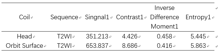
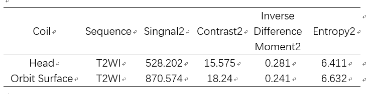
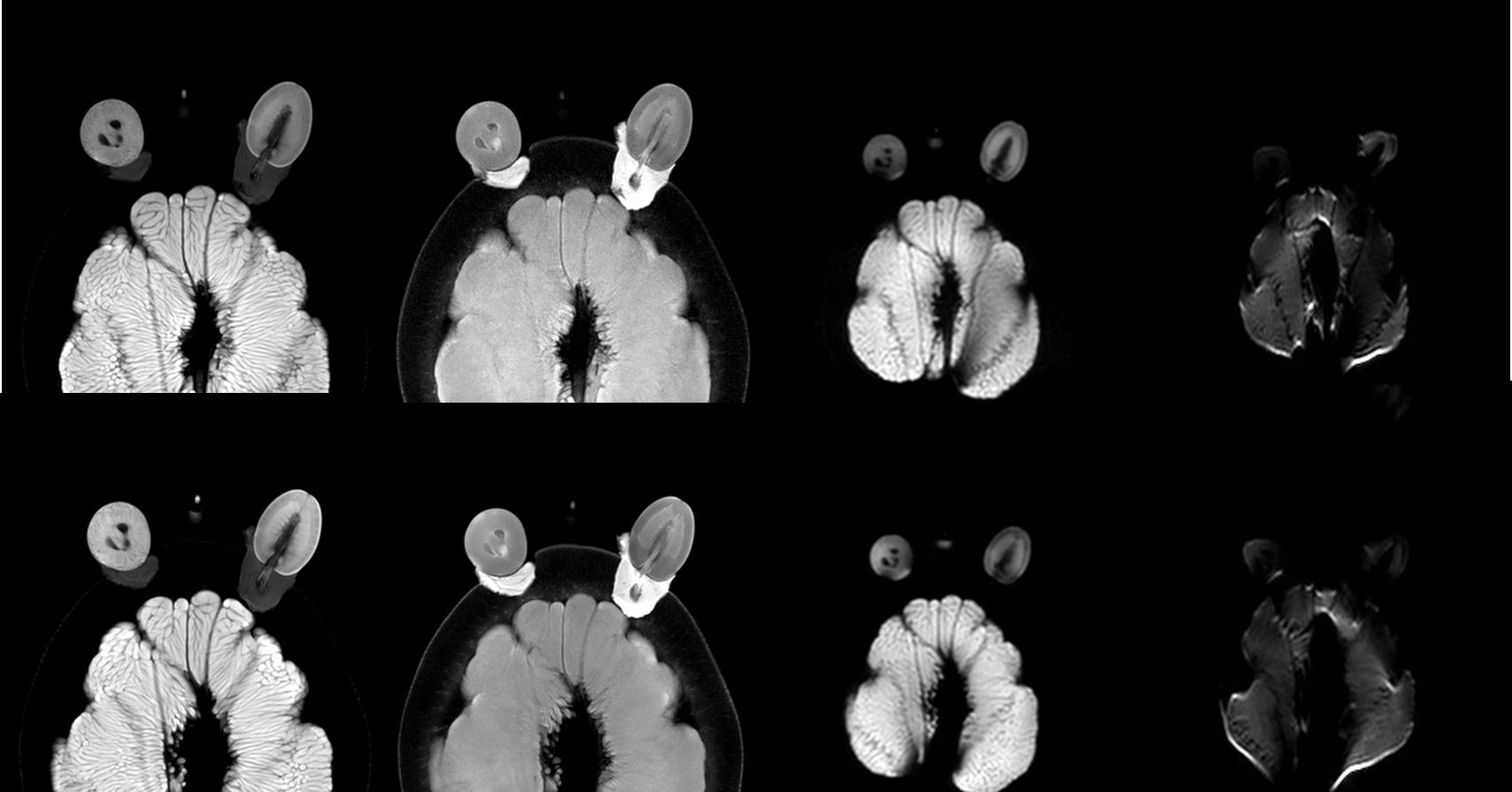
A fruit phantom was designed for this study. Fresh date and cherry tomato were placed in digged holes in peel of grapefruit; grape stems wrapped with lard was used to fill the spaces and connect date and cherry to the grapefruit. Imaging was performed using the new custom-made orbit surface coil on a 3T MR system (Ingenia 3T, Philips Medical Systems); another scan was performed with standard 15-channel head coil (T2WI-mDixon, T1WI-mDixon and DWI-TSE, DWI-EPI). Signal Intensity, Contrast, Inverse Difference Moment and Entropy of two datasets from normal and new coils were measured in Software ImageJ(https://imagej.nih.gov/ij/) via the texture analysis tool. All the image qualities and measurements via ImageJ were compared by two experienced radiologists.Results and discussion
DWI-EPI imaging was challenging due to image distortions from susceptibility artifacts in the fruit phantom in both coils, which showed a good mimic to the true anatomy. The custom-made orbit surface coil can acquire significantly higher signal at cherry tomato (simulated eye and optic nerve) and grapefruit (simulated intracranial structures including cavernous and intracranial optic nerve) than standard head coil with the same clinical parameters. For T2WI images, the contrast and entropy values from new orbit surface coil are higher than that of standard head coil while the inverse difference moment from new coil is lower than that of standard head coil (Table1 and Table 2). In other words, the new coil can depict more texture during T2 Weighted Imaging. However, the subjective impression of grapefruit T2WI images in new surface coil is not as good as standard head coil. Besides, the texture analysis showed that the new surface coil had little advantages over the standard head coil in T1WI and DWI-TSE images.Conclusion
The new custom-made orbit surface coil generated images higher signal and better depict the heterogeneity of the phantom structure. Particularly it showed advantages in cherry tomato (simulated eye and optic nerve), while we observed less image contrast in deeper grapefruit (simulated intracranial structures including cavernous and intracranial optic nerve) which is the limit of this coil due to the elements’ geometry design.
There were mismatches between images acquired by orbit surface coil and standard head coil. Further research is needed to explain the inconsistency between objective results and subjective impression of grapefruit images.
Acknowledgements
NoReferences
[1] Georgouli T, Chang B, Nelson M, et al. Use of high resolution microscopy coil MRI for depicting orbital anatomy. Orbit, 2008, 27(2): 107-114.
[2] Qinghua Chen,et al, Study of optimizing MRI protocols for eyeball. Chin J Magn Reson Imaging,2012, 3( 5):324-330.
[3] Kang LL, Yu XE. Analysis of the influencing factors on signal-to-noise ratio of MR images. Radiol Practice, 2001, 16(6): 404.
[4] Kang LL, Lu GW. The experiment research on the influence of some MR parameters on signal-to-noise ratio. J Practic Radiol, 2003, 19(11): 971-973.
[5] Guo Y, Zhang JW, Zhang XD. The improvement of signalto-noise ratio in magnetic resonance image. J Biom Engineering, 2002, 19(3): 493-495.
Figures