5412
How to separate peripheral nerves from musclesIbrahim Ibrahim1, Jaroslav Tintěra1, Vít Herynek1, Antonín Škoch1,2, Ivan Humhej3, and Milan Hájek1
1Department of Diagnostic and Interventional Radiology, Institute for Clinical and Experimental Medicine (IKEM), Prague, Czech Republic, 2Institute for Clinical and Experimental Medicine (IKEM), Prague, Czech Republic, 3Department of Neurosurgery, Masaryk Hospital, Usti nad Labem, Czech Republic
Synopsis
MR tractography of the peripheral nerves (PN) is challenging due to the difficulty to acquire high quality DWI data for peripheral nerve bundles reconstruction. The aim of this study was to propose an algorithm for separation LSP bundles from muscles using segmentaion of cauda equina and normalized quantitative anisotropy.
Purpose/Introduction
MR tractography of the peripheral nerves (PN) is challenging due to the difficulty to acquire high quality DWI data for peripheral nerve bundles reconstruction. Notably challenging is the separation of the lumbosacral (LSP) bundles from musculoskeletal fibers as both structures have similar fractional anisotropy values (1-2). As a consequence, muscles often contaminate calculated PN fibers that in turn exhibit as rather thick bundles. The aim of this study was to propose an algorithm for separation LSP bundles from muscles using normalized quantitative fractional anisotropy (NQA) instead fractional anisotropy (FA).Methods
five healthy volunteers (2 females, 3 males, mean age of 29.3 ± 5.6 years, range 19-36 years) underwent MRI examinations in the supine position on a 3T MR scanner using a 12-channel phased-array body coil with the following optimized measurement protocol: 1) Diffusion-weighted images obtained by the spin-echo echo-planar imaging (SE-EPI) sequence with the acquisition parameters: voxel size of 3×3×3 mm3, TR/TE = 11100/79 ms, 100 axial slices, number of diffusion directions 30, two b values: 0 and 700 s/mm2 and total acquisition time of 12:03 min. 2) Coronal T2 weighted 3D STIR (short-term inversion recovery) SPACE (sampling perfection with application optimized contrast using varying flip angle evaluation) sequence used for high-resolution MR neurography with the voxel size of 1×1×1 mm3, TR/TE = 2000/149 ms, TI = 160 ms and total acquisition time of 10:00 min. The DTI data were firstly corrected for distortions and eddy current effects using an eddy correct tool in the FSL software (http://fsl.fmrib.ox.ac.uk/fsl/fslwiki/). Afterwards, the data were reconstructed using the DSI studio (http://dsi-studio.labsolver.org/) with the Generalized Q-sampling Imaging - GQI algorithm. Cauda equina (CE) was segmented from the corrected b = 0 images using the ITK-SNAP Medical Image Segmentation Tool (http://sourceforge.net/projects/itk-snap/). Thereafter, the segmented CE was used as a set of seed points with three additional regions of interest (ROIs), placed along the left L3 nerve pathway proxmally, medially and distally with the following tracking parameters: NQA was set to 0.02, the angular threshold 30º. ROIs in contralateral side were placed symmetrically for reconstruction of the right L3 nerve. The above steps were repeated for the reconstruction of the remaining lumbosacral plexus bundles (Fig. 1c). MRT of the psoas major muscle (PMM) was also performed using combination of ROIs (Fig. 2). All regions of interest were manually selected at different levels of the spinal cord nerves and PMM muscles using normalized quantitative anisotropy (NQA) images, orientation distribution function (ODF) maps and T2 3D STIR SPACE images. Mean values of FA, NQA and ADC of the studied nerve bundles were calculated in each subject for PLS bundles (L3-S2, Fig. 1) and also in the PMM (Fig. 2). Fractional anisotropy, apparent diffusion coefficient and normalized quantitative anisotropy in the LSP nerves and the PMM were statistically compared. As the data sets were small and did not have normal distribution, a nonparametric Mann-Whitney U test was employed. p < 0.05 was considered to indicate a statistically significant difference. The result of MR tractography of the LSP and PMM reconstruction is shown in Fig. 1 and Fig. 2.Results
The mean diffusion indices of the PLS/PMM were FA (0.27±0.02/0.23±0.02), ADC (1.89±0.14/1.10±0.16) and NQA(0.19±0.06/0.07±0.02). Difference in FA in the LSP nerves and the PMM was not significant, whereas ADC and NQA significantly differed between LSP nerves and the PMM (both p=0.012).Conclusion
Our results show similar fractional anisotropy values of the lumbosacral bundles and psoas major mascles. MR tractography of the LSP bundles reconstruction can be optimized in terms of eliminating the muscle fibers contamination by using normalized quantitative anisotropy instead fractional anisotropy.Acknowledgements
The study was supported by Ministry of Health of the Czech Republic, grant No. 17-28587A and MHCZ-DRO 00023001IKEM.References
1. Longwei X (2012) Clinical application of diffusion tensor magnetic resonance imaging in skeletal muscle. Muscles Ligaments Tendons J 2:19-24.
2. Van der Jagt PK, Dik P, Froeling M, Kwee TC, Nievelstein RA, ten Haken B, Leemans A (2012) Architectural configuration and microstructural properties of the sacral plexus: a diffusion tensor MRI and fiber tractography study. Neuroimage 62:1792-1799.
Figures
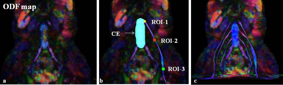
Fig.
1.
MR tractography of the lumbosacral plexus. a) orrientation disstribution function
(ODF) map. b) segmented cauda equina was used as a set of seed points to
reconstruct the major pathway roots of lumbosacral bundles. Three additional
regions of interest (ROIs) were placed
along the left L3 nerve pathway proxmally, medially and distally with the
following tracking parameters: NQA was set to 0.02, the angular
threshold 30º, the step size 1.5 mm and primary orientation were setin order to
visualize the left L3 nerve. The above steps were repeated for the
reconstruction of the remaining lumbosacral plexus bundles (c).
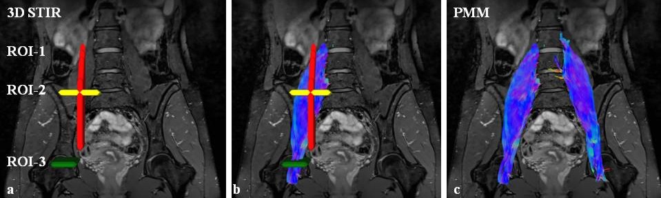
Fig. 2: A representative sample reconstruction of the
psoas major muscle (PMM) using three regions of interest (ROIs). One ROI was placed at the sagittal
plane (red) and two ROIs in the transversal plane (ROI-2 (yellow) in the L5/S1
level and ROI-3 (green) in the head level of the femur (a). MR tractography of the
PMM is shown in b and c. Cauda equina was also used as a region of
avoidance to eliminate lumbosacral
plexus fiber bundles.