5410
Correlation of tumor blood flow in head and neck squamous cell carcinoma by pseudo-continuous arterial spin labeling with parameters of dynamic contrast-enhanced MRI1Asan Medical Center, Seoul, Republic of Korea, 2Siemens healthcare, MR Application development, Erlangen, Germany
Synopsis
The aim of this prospective study was to evaluate the correlation between tumor blood flow (TBF) measurement using pseudo-continuous arterial spin labeling (pCASL) and parameters of DCE-MRI in patients with head and neck squamous cell carcinoma (HNSCC). We scanned 26 patients with HNSCC using 3T MRI with both pCASL and DCE-MRI. There were significant correlation between TBF of pCASL and wash-in, signal enhancement ratio, and Vp of DCE-MRI, with a correlation coefficient of 0.649, 0.642, and 0.507, respectively (P<0.01). The pCASL can be a useful tool for noninvasive assessments of the TBF in patients with HNSCC.
INTRODUCTION
The recent use of the arterial spin labeling (ASL) technique has allowed the noninvasive measurement of the tissue blood flow even in head and neck lesions. For head and neck squamous cell carcinomas (HNSCCs), several studies have demonstrated that tumor blood flow (TBF) are important biological factors for the diagnosis and treatment, respectively. The relationship between parameters of dynamic contrast-enhanced MRI (DCE-MRI) and TBF of ASL has been unclear, and a few studies have investigated this relationship. The aim of this prospective study was to evaluate the feasibility of TBF measurement using pCASL, in comparison to parameters of DCE-MRI in patients with HNSCC.METHODS
This prospective study protocol was reviewed and approved by the hospital review board, and the requirement for informed consent for data evaluation was waived. Written informed consent to undergo the MRI protocol was obtained from all patients before each examination. The methods and reporting of results are in accordance with the STROBE (Strengthening the Reporting of Observational Studies in Epidemiology) statement.
Study patients The study population was obtained from a prospective cohort of consecutive patients who were newly diagnosed with HNSCC by pathologic examination with no prior treatment and underwent pretreatment MRI with at a 2700-bed academic tertiary referral hospital, between June 2016 and February 2017. Patients were excluded if they had any of following criteria: younger than 18 years of age: suboptimal image quality due to motion or small lesion.
MRI protocol
DCE-MRI examinations were performed using a 3-T scanner (Skyra, Siemens Healthcare) with a 64-channel neurovascular coil. The dynamic acquisition was performed with a temporal resolution of 4.0 seconds, and contrast material was administered after 11 baseline dynamics (total, 150 dynamics). The detailed imaging parameters for DCE-MRI were as follows: a slice thickness of 4 mm with no gap; 21 slices; z-axis coverage of 84 mm; spatial in-plane resolution of 180 × 180; TR/TE, 4.5/1.9 msec; flip angle, 25°; FOV, 17.8 cm; and total acquisition time of 9 minutes 24 seconds. The acquisition of pCASL was performed by using multi-shot spin-echo echo planar imaging to obtain control and labeled images. The labeling slab was placed just under the bifurcation of the internal and external carotid arteries by using the coronal T2WI as the reference. The pCASL parameters were as follows: labeling duration, 1650 ms; post-label delay, 1330 ms; TR, 4600 ms; TE, 15 ms; flip angle, 180°; number of shots, 1; FOV, 190×190 mm; matrix, 128×128; slice thickness, 3 mm; number of slices, 42; and scanning time, 6 min 2s.
Data analysis
TBF was analyzing using AFNI software. Parameters of DCE-MRI were quantitatively and qualitatively analyzed using dedicated software (Olea Sphere 2.3), based on the extended Tofts model and model-free method. Parameters of DCE-MRI were used to assess tissue and vascular permeability characteristics: Ktrans, Kep, Ve, Vp, wash-in, wash-out, AUC, peak enhancement, and signal enhancement ratio. We drew a volume of interest encompassing the entire tumor volume.
Statistical analysis
We analyzed the correlations between the TBF and parameters of DCE-MRI using Pearson’s correlation coefficients (r < 0.2, poor correlation; r = 0.2–0.4, weak correlation; r = 0.41–0.6, moderate correlation; r = 0.61–0.8, good correlation; r > 0.8, excellent correlation). All statistical analyses were performed using MedCalc for Windows (version 15.0; MedCalc Software, Ostend, Belgium), and p < 0.05 was considered statistically significant.
RESULTS
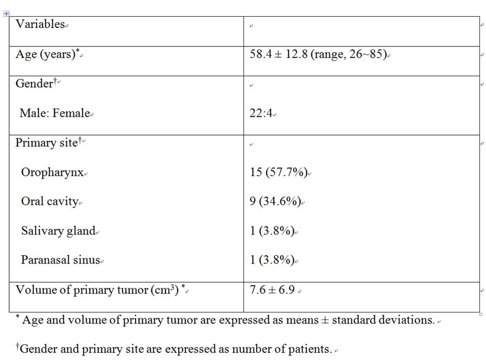
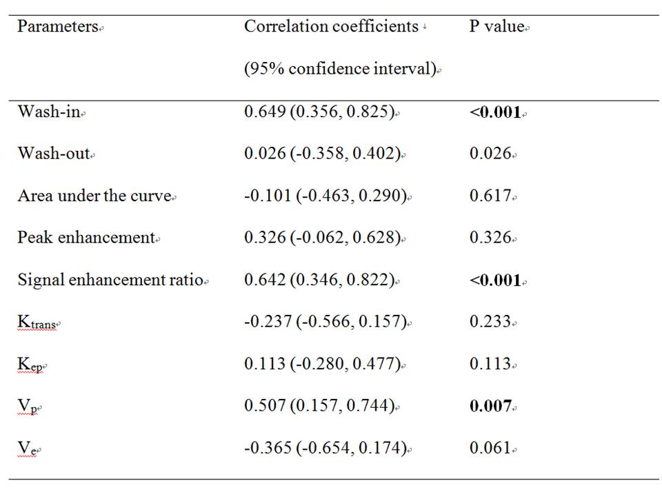
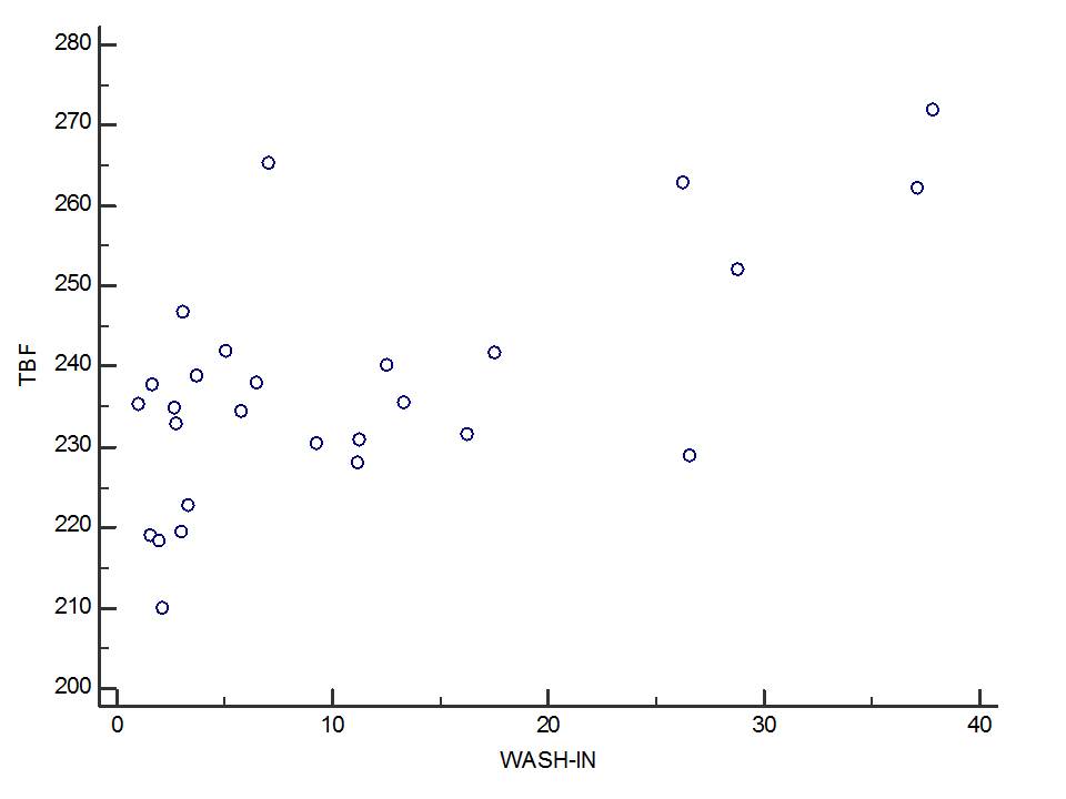
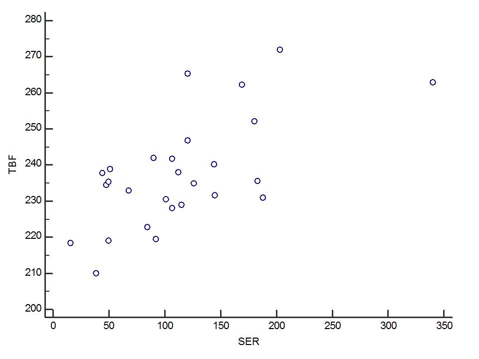
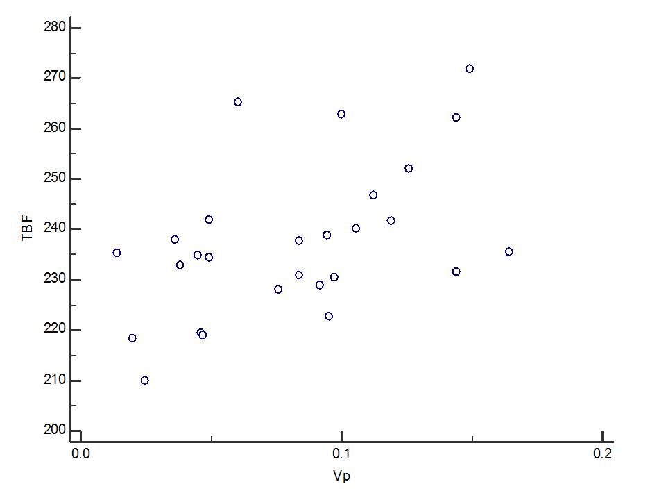
During study period, 29 patients were considered to be eligible. After exclusion of 3 patients who had suboptimal image quality due to motion or small lesion in the mobile tongue, 26 patients were finally enrolled. Tigure 1 shows the demographic data of patients. The most common primary site was oropharynx (n=15, 57.7%) followed by oral cavity (n=9, 34.7%). Among oropharynx, palatine tonsil was 11 patients and base of tongue was 4 patients. There were significant correlation between TBF values measured by pCASL and wash-in, signal enhancement ratio, and Vp measured by DCE-MRI, with a Pearson’s correlation coefficient of 0.649, 0.642, and 0.507, respectively (P<0.01) (Figure 2 and 3).DISCUSSION
Our prospective study demonstrated that there was moderate correlation between TBF values measured by pCASL and parameter of DCE-MRI (wash-in, signal enhancement ratio, and Vp ) in the patients with HNSCC. In our study, a pCASL scan was successfully achieved in 90% (26/29) HNSCC patients.CONCLUSION
In conclusion, pCASL can be a useful tool for noninvasive assessments of the TBF in patients with HNSCC. The TBF values obtained by pCASL will enable evaluations of HNSCC, similar to parameters measured by DCE perfusion.Acknowledgements
noneReferences
1. Editors PM. Observational studies: getting clear about transparency. PLoS Med. 2014;11:e1001711.
2. Fujima N, Sakashita T, Homma A, et al. Glucose Metabolism and Its Complicated Relationship with Tumor Growth and Perfusion in Head and Neck Squamous Cell Carcinoma. PloS one 2016;11(11):e0166236.
3. Viallon M, Cuvinciuc V, Delattre B, et al. State-of-the-art MRI techniques in neuroradiology: principles, pitfalls, and clinical applications. Neuroradiology 2015;57(5):441-67.
4. Grade M, Hernandez Tamames JA, Pizzini FB, Achten E, Golay X, Smits M. A neuroradiologist's guide to arterial spin labeling MRI in clinical practice. Neuroradiology 2015;57(12):1181-202.
5. Alsop DC, Detre JA, Golay X, et al. Recommended implementation of arterial spin-labeled perfusion MRI for clinical applications: A consensus of the ISMRM perfusion study group and the European consortium for ASL in dementia. Magn Reson Med 2015;73(1):102-16.
6. Griffith B, Jain R. Perfusion Imaging in Neuro-Oncology: Basic Techniques and Clinical Applications. Magn Reson Imaging Clin N Am 2016;24(4):765-779.
7. Telischak NA, Detre JA, Zaharchuk G. Arterial spin labeling MRI: clinical applications in the brain. J Magn Reson Imaging 2015;41(5):1165-80.
8. Fujima N, Kudo K, Yoshida D, et al. Arterial spin labeling to determine tumor viability in head and neck cancer before and after treatment. J Magn Reson Imaging 2014;40(4):920-8.
9. Fujima N, Kudo K, Tsukahara A, et al. Measurement of tumor blood flow in head and neck squamous cell carcinoma by pseudo-continuous arterial spin labeling: comparison with dynamic contrast-enhanced MRI. J Magn Reson Imaging 2015;41(4):983-91.
10. Amukotuwa SA, Yu C, Zaharchuk G. 3D Pseudocontinuous arterial spin labeling in routine clinical practice: A review of clinically significant artifacts. J Magn Reson Imaging 2016;43(1):11-27.
11. Kato H, Kanematsu M, Watanabe H, et al. Perfusion imaging of parotid gland tumours: usefulness of arterial spin labeling for differentiating Warthin’s tumours. European Radiology 2015;25(11):3247-3254.
12. Fujima N, Kameda H, Tsukahara A, et al. Diagnostic value of tumor blood flow and its histogram analysis obtained with pCASL to differentiate sinonasal malignant lymphoma from squamous cell carcinoma. Eur J Radiol 2015;84(11):2187-93.
13. Fujima N, Nakamaru Y, Sakashita T, et al. Differentiation of squamous cell carcinoma and inverted papilloma using non-invasive MR perfusion imaging. Dento maxillo facial radiology 2015;44(9):20150074.
14. Fujima N, Yoshida D, Sakashita T, et al. Usefulness of Pseudocontinuous Arterial Spin-Labeling for the Assessment of Patients with Head and Neck Squamous Cell Carcinoma by Measuring Tumor Blood Flow in the Pretreatment and Early Treatment Period. AJNR Am J Neuroradiol 2016;37(2):342-8.
Figures