Takuya Aoike1, Noriyuki Fujima2, Masami Yoneyama3, Kinya Ishizaka1, Hiroyuki Sugimori4, and Kohsuke Kudo2
1Department of Radiological Technology, Hokkaido University Hospital, Sapporo, Japan, 2Department of Diagnostic and Interventional Radiology, Hokkaido University Hospital, Sapporo, Japan, 3Philips Healthcare, Tokyo, Japan, 4Faculty of Health Sciences, Hokkaido University, Sapporo, Japan
Synopsis
We assessed the rapid acquisition design in 3D-MR neurography (3D-MRN) using
compressed sensing (CS) with the combination of the parallel imaging
technique. High sparsity in 3D-MRN raw data was considered to be
compatible with high CS acceleration factor. This result will be
make patients comfortable in daily clinical MRN scanning.
Purpose
3D-magnetic resonance neurography (3D-MRN) of brachial
plexus can easily obtain the anatomical nerve shape in greater detail; however,
this technique typically requires long acquisition time such as 5-6 minutes1.
Compressed sensing (CS) is described one of popular fast data-acquisition method
using the sparsity of the target data set2. Because 3D-MRN mainly
consists of bright peripheral nerve signal and suppressed background signal
only, the 3D-MRN is expected to have a lot of sparsity in its raw-data. Therefore,
the addition of CS technique to the conventional 3D-MRN acquisition is
considered to be useful for short acquisition time. The aim of this study was
to develop the rapid acquisition design in 3D-MRN using CS with the combination
of the parallel imaging technique.Methods
All
scanning was performed using a 3.0 Tesla scanner (Achieva TX; Philips
Healthcare, Best, Netherlands) with a 16-channel neurovascular coil. MRN used
motion-sensitized driven-equilibrium (MSDE) turbo spin echo (TSE) sequence
with: TR, 2200 ms; TE, 170 ms; FOV, 300×300 mm; Voxel size, 1.17×1.18×2.00 mm;
Slice thickness 2 mm×170 slices. As a post-processing, 3D-coronal view was
reconstructed by using maximum intensity projection (MIP) algorithm. For the
image acceleration, we used the combined acceleration factor defined as CS-SENSE factor which
means total acceleration with the combination of CS and conventional parallel
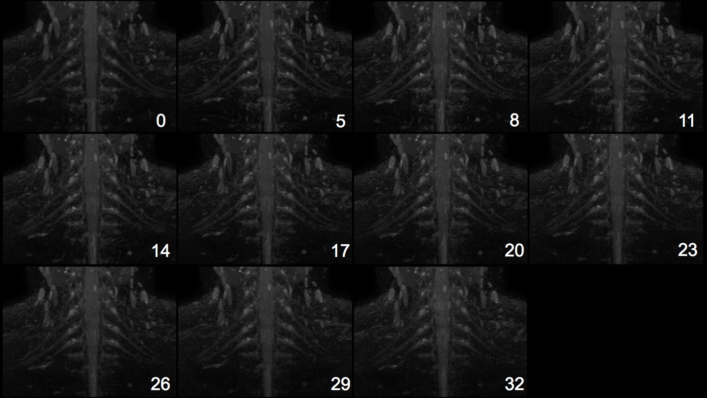
imaging acceleration factor. CS-SENSE factor of 0, 5, 8, 11, 14, 17, 20,
23, 26, 29 and 32 were respectively used. Each scanning time of various
CS-SENSE factors were as follows; CS-SENSE factor of 0 (6’32), 5 (5’28), 8 (3’27),
11 (2’32), 14 (1’59), 17 (1’39), 20 (1’24), 23 (1’13), 26 (1’06), 29 (0’57), 32
(0’53). The image quality of the depicted brachial plexus was visually
evaluated based on the degree of depiction of the nerve sheath compared to the
background signal using the following 3-grading system: 1, poor; 2, moderate;
3, good visibility of the nerve sheath by five experienced radiological technologists. In
statistical analysis, the obtained score was compared between various CS-SENSE
factors with multiple comparison of Dunnett's test with the reference data of
CS-SENSE factor of 0. Statistical significance was set to p<0.01.
Results
Coronal MIP images of 3D-MRN with various
CS-SENSE factors were shown in Fig.1.
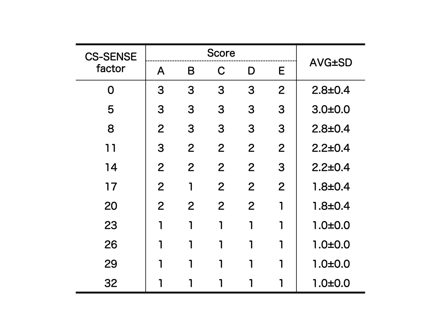
In visual evaluation, visual score in
3-grading system was not significantly different in CS-SENSE factor of 5 (3.0±0.0), 8 (2.8±0.4), 11 (2.2±0.4) and 14 (2.2±0.4) compared to the reference data of
CS-SENSE factor 0 (2.8±0.4). In contrast, CS-SENSE factor of 17 (1.8
±
0.4), 20 (1.8 ± 0.4), 23 (1.0±0.0), 26 (1.0±0.0), 29 (1.0±0.0) and 32 (1.0±0.0) were significantly different compared
to CS-SENSE factor of 0 (p<0.01).
Representative case was presented in Table.1.Discussion and Conclusion
From the results, high sparsity in 3D-MRN raw data
was considered to be compatible with high CS acceleration factor. Especially,
only 1’59 minutes scanning with CS-SENSE factor of 14 was visually acceptable
compared to conventional 5-6 minutes scanning. Such rapid MRN scanning will make
patients comfortable in daily clinical MR scanning.Acknowledgements
No acknowledgements.References
1. Yoneyama M, Takahara T, Kwee TC, et al. Rapid High Resolution MR Neurography with a Diffusion-weighted Pre-pulse. Magn Reson Med Sci. 2013;12(2):111-9
2.
Lustig M, Donoho D, Pauly JM.
Sparse MRI: The Application of Compressed Sensing
for Rapid MR Imaging. Magn Reson Med. 2007 Dec;58(6):1182-95