5390
The Effect of Respiration on Apparent Diffusion Coefficient of the Brain1Institute of Medical, Pharmaceutical and Health Sciences, Kanazawa University, Kanazawa, Japan, 2Department of Neurosurgery, Nagoya City University, Nagoya, Japan
Synopsis
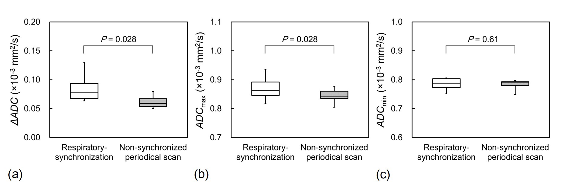
ADC of the brain significantly changes during the cardiac cycle because of intracranial pressure changes and fluid fluctuations. We hypothesized that ADC of the brain changes during the respiratory cycle because intracranial pressure changes during the respiratory cycle. We determined the maximum and minimum ADC (ADCmax and ADCmin) and maximum change in ADC (ΔADC) in the respiratory cycle and non-synchronized periodical data. ADCmax and ΔADC in the white matter with respiratory-synchronization were significantly greater than those with non-synchronized periodical scan, whereas there was no significant difference in ADCmin between the scans. ADC of the brain changes during the respiratory cycle.
Introduction
Apparent diffusion coefficient (ADC) of the brain has been widely used for the quantitative evaluation of various brain diseases. We have previously reported that ADC of the brain significantly changed during the cardiac cycle because of intracranial pressure changes and fluid fluctuations, even with minimization of the bulk motion effect.1-2 Since intracranial pressure changes during the respiratory cycle,3 we hypothesized that ADC of the brain also changes during the respiratory cycle. Therefore, we assessed dynamic changes in ADC of the brain during the respiratory cycle using respiratory-synchronized cine diffusion magnetic resonance imaging (MRI).Materials and Methods
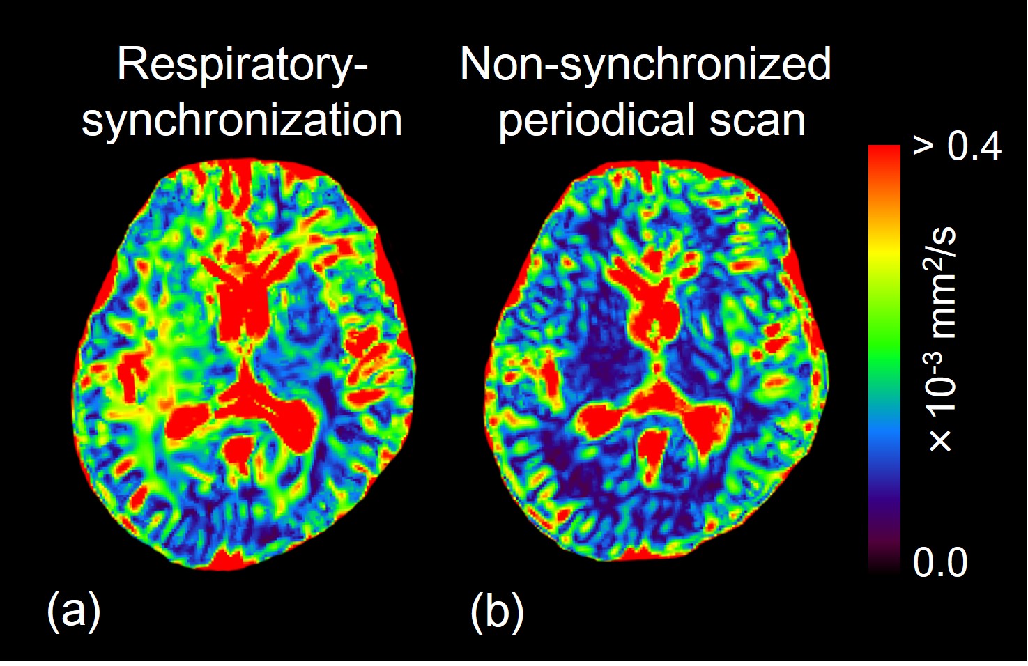
On a 3.0-T MRI system, we performed single-shot diffusion echo-planar imaging with respiratory-synchronization and non-synchronized periodical scan (at random). We obtained ADC images in healthy male subjects using the following imaging parameters: repetition time, one respiratory cycle; echo time, 65 ms; imaging matrix, 64 × 64; field of view, 256 mm; slice thickness, 4 mm; b-value, 0 and 1000 s/mm2; number of signals averaged, 2; number of phases, 14; parallel imaging factor, 2; and half scan factor, 0.6. Then, we placed regions of interest in the white matter and determined the maximum and minimum ADC (ADCmax and ADCmin, respectively) and maximum change in ADC (ΔADC) in both the respiratory cycle and non-synchronized periodical data.Results and Discussion
ΔADC and ADCmax in the white matter obtained with respiratory-synchronization were significantly greater than those with non-synchronized periodical scan, whereas there was no significant difference in ADCmin between the scans (Figs. 1 and 2). These results indicate that ADC of the brain is affected by respiration.Conclusion
ADC of the brain changes during the respiratory cycle, and analysis of the dynamic ADC change makes it possible to obtain more accurate ADC.Acknowledgements
No acknowledgement found.References
1. Ohno N, Miyati T, Mase M, et al. Idiopathic normal-pressure hydrocephalus: temporal changes in ADC during cardiac cycle. Radiology. 2011; 261: 560-565.
2. Takahashi Y, Hori M, Shimoji K, et al. Changes in delta ADC reflect intracranial pressure changes in craniosynostosis. Acta Radiol Open. 2017; 6: 2058460117728535.
3. Yildiz S, Thyagaraj S, Jin N, et al. Quantifying the influence of respiration and cardiac pulsations on cerebrospinal fluid dynamics using real-time phase-contrast MRI. J Magn Reson Imaging. 2017; 46: 431-439.
Figures