5385
White matter micro-structural alterations in Hypothyroid: A Diffusion Kurtosis Imaging studyMukesh Kumar1, Poonam Rana1, Deepak Sharma1, Ratnesh Kanwar2, Tarun Sekhri2, Maria D’souza1, and Subash Khushu1
1NMR Research Center, Institute of Nuclear Medicine and Allied Sciences, New Delhi, India, 2Thyroid Research Center, Institute of Nuclear Medicine and Allied Sciences, New Delhi, India
Synopsis
The aim of our study was to assess changes in brain tissue microstructures in hypothyroid patients using Diffusion Kurtosis Imaging (DKI). The water diffusion in living tissue is hindered by interactions with other molecules and cell membranes. Therefore, water movement in biological tissue is often non-Gaussian and this non-Gaussian behavior may contain useful information related to tissue structure and pathophysiology. Our findings demonstrate widespread reduced kurtosis indices MK, AK; diffusion index FA was decreased and AD was increased in major white matter pathways and such abnormal white matter structure may be linked to cognitive and behavioral impairment in hypothyroid patients.
Introduction
Hypothyroidism is a clinical condition that is characterized by elevated levels of thyroid-stimulating hormone (TSH) and low levels of free tri-iodothyronine (FT3) and free thyroxine (FT4). Thyroid hormones (THs) play a key role in regulating neuronal migration, neuronal differentiation, and dendritic proliferation during mammalian cerebellum development and maturation 1. The deficiency of THs during human brain development leads to irreversible mental retardation and neurological deficits 2. An adulthood overt hypothyroidism is associated with neuropsychiatric complaints and symptoms 3. In order to assess changes in brain tissue micro-structure in hypothyroid patients, we used Diffusion Kurtosis Imaging (DKI). The water diffusion in living tissue is hindered by interactions with other molecules and cell membranes. Therefore, water movement in biological tissue is often non-Gaussian and this non-Gaussian behavior may contain useful information related to tissue structure and pathophysiology 4. Magnetic Resonance DKI is an emerging technique with the potential to quantify properties of tissue micro-structures that may not be observable using conventional Diffusion Tensor Imaging (DTI).Materials and methods
22 control subjects (mean age ± SD = 31.75 ± 8.60) and 20 hypothyroid patients (mean age ± SD = 31.94 ± 10.49) participated in the study. The recruited hypothyroid patients had decrease FT4 and FT3 levels and TSH (>10 μIU/ml). The informed consent was obtained from all the subjects prior to DKI study. None of the subjects had any history of neurological or psychiatric disorders. The study was approved by the institutional ethics committee. MR Imaging was accomplished using a 3-Tesla MRI scanner (Magnetom, Skyra, Siemens) with a 20 channel head and neck coil and 25 mT/m actively shielded gradient system. The conventional MR imaging was done prior to DKI to rule out any structural abnormality using routine T2-weighted turbo spin-echo sequence. DKI data were acquired using a single-shot echo-planar dual SE sequence in 30 directions with ramp sampling. Diffusion-weighted acquisition parameters were: 5 b-value= 0,500,1000,1500 and 2000 s/mm2 , slice thickness=4.5 mm with no inter-slice space, number of slices=30, FOV=230 mm×230 mm, matrix size = 128 × 128, spatial resolution = 1.797 mm X 1.797 mm X 4.5mm, flip angle 90°, TR = 7300 ms, TE = 138 ms and NEX=2. Eddy current-induced distortion, motion artifacts, and skull striping were done using FMRIB software library (FSL 5.9, http://www.fmrib.ox.ac.uk/fsl). DKI parameters (MK, AK, RK, FA, MD, AD and RD) were calculated using Diffusion Kurtosis Estimator (DKE) (http://www.nitrc.org/projects/dke). DTI parameters (FA, MD, AD and RD) estimated using b = 0 and 1000 s/mm2, because DTI parameters based on the mono-exponential model on a single non-zero b value in six independent directions. Post-processing performed with the help of Tract-Based Spatial Statistics (TBSS) using FMRIB software library (FSL, http://www.fmrib.ox.ac.uk/fsl).Results and Discussion
Voxelwise analysis revealed a significant decrease in kurtosis parameters Axial Kurtosis (AK), Mean Kurtosis (MK) in hypothyroid patients as compared to healthy controls. Also, our result showed significantly decreased diffusion parameter Fractional Anisotropy (FA) and significantly increased Axial Diffusion (AD) in hypothyroid patients as compared to healthy controls (See Figure 1, Table 1). The decreased kurtosis parameter MK and AK suggest the loss of cellular structure in hypothyroid patients 4. Whereas decreased FA and increased AD in hypothyroid patients may be suggested edema, demyelination and structural disintegration of white matter fiber tract in hypothyroid patients 5,6. Our findings suggest that deterioration of these white matter fiber tracts in hypothyroidism patients may contribute to underlying dysfunction in cognitive functions.Conclusion
Our findings suggest that hypothyroid subjects demonstrate widespread reduced kurtosis indices MK, AK; decrease diffusion index FA and increased index AD in major white matter pathways and such abnormal white matter structure may be linked to some cognitive and behavioral impairment. However, the correlation among white matter fiber strength, clinical parameters and neuropsychological scores of subjects needs to be studied.Acknowledgements
We acknowledge that this research was supported by 'Defense Research and Development Organization' (DRDO), Ministry of Defense, Government of India.References
- Diez D, Grijota-Martinez C, Agretti P, De Marco G, Tonacchera M, Pinchera A, Morreale de Escobar G, Bernal J, Morte B. Thyroid hormone action in the adult brain: gene expression profiling of the effects of single and multiple doses of triiodo-L-thyronine in the rat striatum. Endocrinology. 2008;149(8):3989-4000.
- Anderson GW. Thyroid hormone and cerebellar development. The Cerebellum. 2008;7(1):60-74.
- Samuels MH, Schuff KG, Carlson NE, Carello P, Janowsky JS. Health status, mood, and cognition in experimentally induced subclinical hypothyroidism. The Journal of Clinical Endocrinology & Metabolism.;92(7):2545-51.
- Zhu J, Zhuo C, Qin W, Wang D, Ma X, Zhou Y, Yu C. Performances of diffusion kurtosis imaging and diffusion tensor imaging in detecting white matter abnormality in schizophrenia. NeuroImage: Clinical. 2015;7:170-6.
- Lu H, Jensen JH, Ramani A, Helpern JA. Three‐dimensional characterization of non‐gaussian water diffusion in humans using diffusion kurtosis imaging. NMR in Biomedicine. 2006 Apr 1;19(2):236-47. 6. Steven AJ, Zhuo J, Melhem ER. Diffusion kurtosis imaging: an emerging technique for evaluating the microstructural environment of the brain. American journal of roentgenology. 2014;202(1):W26-33.
Figures
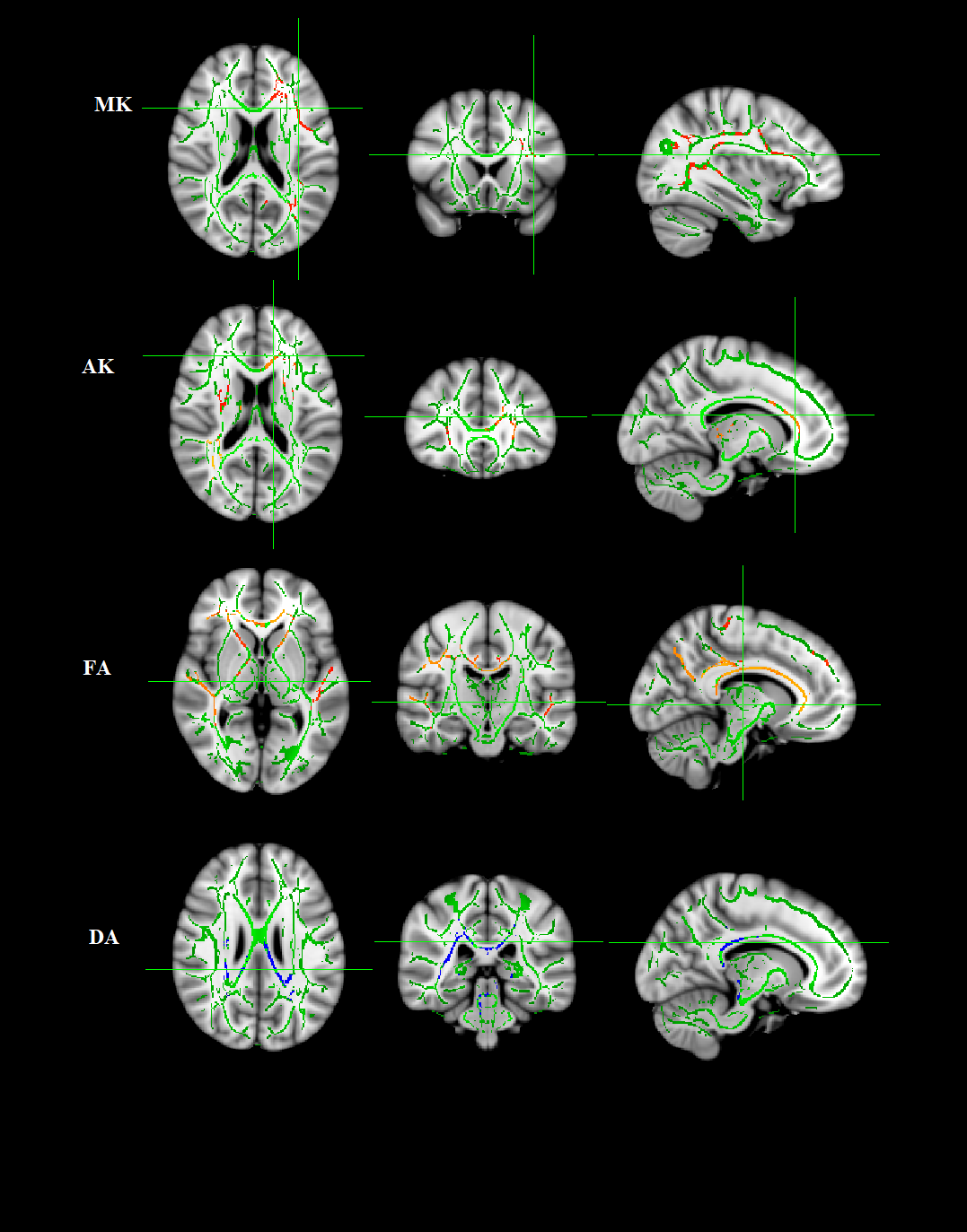
TBSS shows white matter regions with significant
differences in the Mean kurtosis (MK), Axial kurtosis (AK), Fractional
anisotropy (FA) and Axial diffusivity (AD)
between hypothyroid patients and healthy controls (P < .05, FWE corrected). Green
color represents mean FA skeleton of all participants; red yellow color denotes
decrease and blue color represents increased in hypothyroid patients.
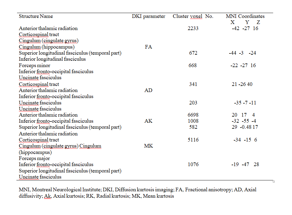
Brain regions showing significant differences in
DKI parameter in control vs. hypothyroid patients in white matter fiber tract