5375
Oscillating Gradient Spin Echo Diffusion Tensor MRI of Acute Human Stroke1Biomedical Engineering, University of Alberta, Edmonton, AB, Canada, 2Neurology, University of Alberta, Edmonton, AB, Canada, 3Radiology, University of Alberta, Edmonton, AB, Canada
Synopsis
Different spatial scales of micro-structural alteration with disease can be interrogated by varying the time water has to interact with its surroundings (e.g. membranes, etc). Oscillating gradient spin echo (OGSE) diffusion MRI enables much shorter diffusion times (e.g. 6 ms) than typical pulsed gradient spin-echo (PGSE) (e.g. 40 ms). The large diffusion reduction typically observed with PGSE in ischemic lesions in human stroke was markedly less with OGSE, and the OGSE-PGSE difference was greatest for radial diffusivity in ischemic white matter. This result is consistent with the swelling and beading of the axons underlying diffusion contrast in cerebral ischemia.
Introduction
Diffusion-weighted imaging of acute stroke is used diagnostically daily, but despite being discovered 27 years ago there are still uncertainties in the origin of the reduced mean diffusivity (MD). Different spatial scales of micro-structural alteration with disease can be interrogated by varying the time water has to interact with its surroundings (e.g. membranes, etc). Oscillating gradient spin echo (OGSE) diffusion MRI enables much shorter diffusion times (e.g. 4 ms) than the typical pulsed gradient spin-echo (PGSE) (e.g. 40 ms). Ischemic brain still shows reduced diffusion with OGSE, but by not nearly as much as with PGSE in post-mortem rat1 and mouse hypoxia-ischemia2. The implementation of OGSE for human MRI is challenging. The only human stroke OGSE study used a frequency of 50 Hz (i.e. ~ 4 ms diffusion time) and very low b=300 s/mm2 on a 4.7T MRI to show only 8% MD reduction in the white matter of human stroke, as opposed to the typical 37% MD reduction with PGSE3. The purpose here was to evaluate OGSE of acute human stroke on a typical 3T MRI.Methods
Both OGSE and PGSE were acquired with single shot spin-echo EPI on a 3T Siemens Prisma (Advanced Diffusion WIP) with: 20 3 mm axial slices, 1.7x1.7 mm2 resolution, GRAPPA R=2, TR 5600 ms, TE 78 ms, b=500 s/mm2 (higher than in reference [3]), 6 directions, 3 averages, and scan time 2:26 min. The OGSE used trapezoid cosines with 40 Hz (i.e. ~ 6 ms diffusion time, longer than in reference [3]) while PGSE had a diffusion time of 40 ms. The stroke patient characteristics were: Patient 1 - 57 year female, right lacunar stroke in white matter, 0.4 cc lesion volume, scan 55 hours post-onset, NIHSSS 0; Patient 2 - 58 year male, right MCA stroke mostly in gray matter, 9.9 cc lesion volume, scan 19 hours post-onset, NIHSSS 4. DTI was fit with ExploreDTI and custom software was used for ROI measurements to compare OGSE and PGSE-derived DTI metrics in well-defined white matter regions either within the lesion or the corresponding contralateral hemisphere.Results
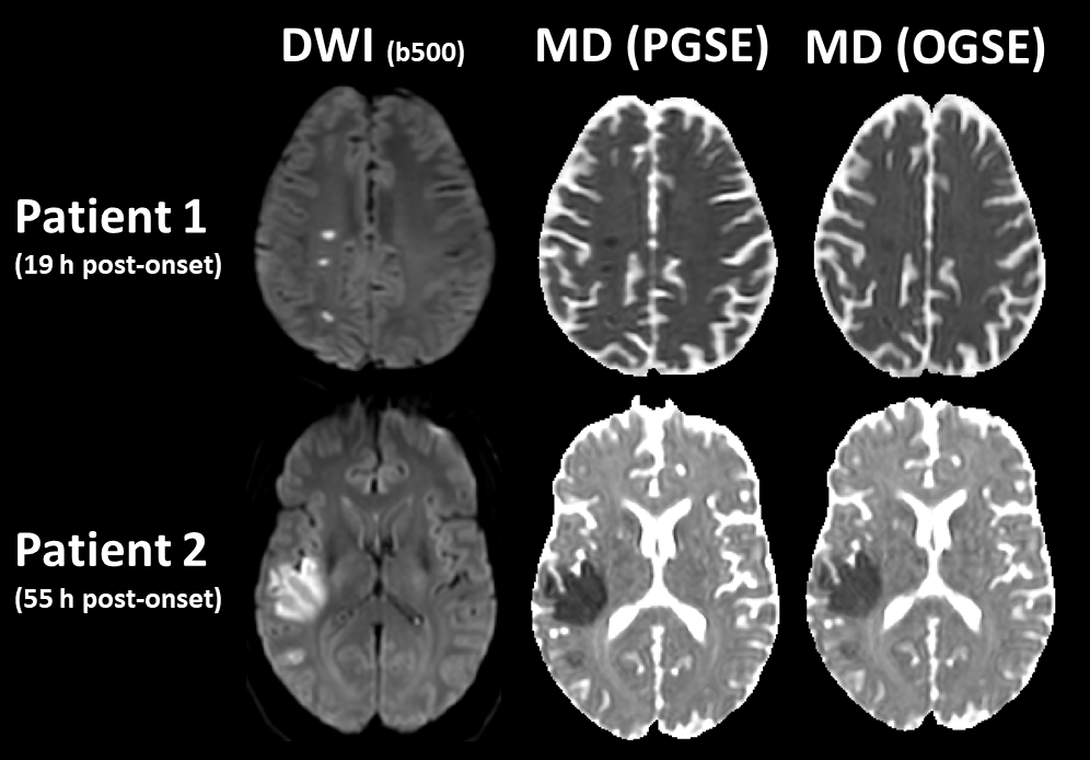
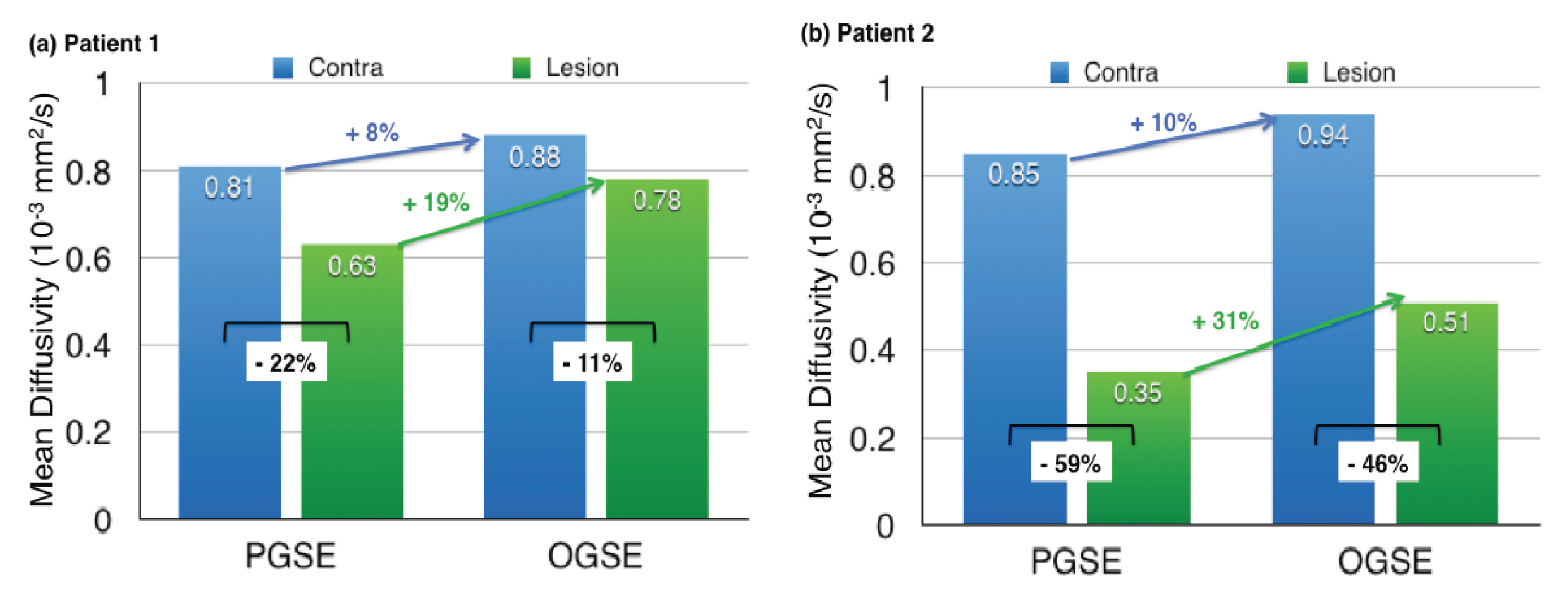
There was diminished stroke lesion contrast on the OGSE MD maps relative to the PGSE MD maps (Figure 1). A very interesting observation in the larger stroke of Patient 2 was that MD within the lesion was lower in white matter than gray matter as measured by PGSE, but this gray/white MD difference was attenuated on OGSE. Thus the effect of microstructure alteration on diffusion becomes more similar for gray and white matter when querying small spatial scales of diffusion. Quantitative MD analyses for both patients are shown in Figure 2. Relative to the contralateral side, MD was reduced in the lesion to a greater extent with PGSE than with OGSE {Patient 1 (P1): -22% drop PGSE vs -11% drop OGSE; Patient 2 (P2): -59% drop PGSE vs -46% drop OGSE}. OGSE yields 8%(P1) and 10%(P2) greater MD than PGSE in contra-lateral white matter (in agreement with reference [4]), but this increase is much greater in the lesion white matter at 19%(P1) and 31%(P2). OGSE also yields greater radial diffusivity than PGSE in contra-lateral white matter {12%(P1) and 17%(P2)}, and this increase is greater than that of axial diffusivity {3%(P1) and 4%(P2)}. Again, OGSE-PGSE differences are greater in the stroke lesion {radial 23%(P1) and 41%(P2) increase versus axial 14%(P1) and 24%(P2) increase}.Discussion and Conclusions
The restrictive effects on water diffusion in an ischemic stroke lesion are less apparent at the short diffusion times enabled by OGSE. This is consistent with previous animal models1,2 and the only other human study3. Relative to reference [3], the current study used a more standard field strength of 3T (versus 4.7T), and a higher b value of 500 s/mm2 (versus 300 s/mm2) to better measure the DTI metrics, but a lower frequency of 40 Hz (versus 50 Hz), and a longer diffusion time of 6 ms (versus 4 ms). The longer diffusion times in this study are consistent with smaller OGSE/PGSE differences than in reference [3]. Greater radial than axial diffusion increase in going from 40 ms (PGSE) to 6 ms (OGSE) diffusion time in ischemic white matter suggests an enlargement of micro-structural space in that dimension. The results of the previous OGSE/PGSE stroke study at 4.7T suggested diffusion contrast in stroke was well explained by axonal swelling and beading3. The result of the current study at 3T with greater b-value also supports this diffusion contrast theory in acute stroke.Acknowledgements
Canada Research Chairs, Heart and Stroke Foundation of Canada, Canadian Institutes of Health Research.References
[1] Does M, Parsons E, Gore J. Oscillating gradient measurements of water diffusion in normal and globally ischemic rat brain. Magn Reson Med 49, 206 (2003).
[2] Wu D, Martin L, Northington F, Zhang J. Oscillating gradient diffusion MRI reveals unique microstructural information in normal and hypoxia-ischemia injured mouse brains. Magn Reson Med 72, 1366 (2014).
[3] Baron C, Kate M, Gioia L, Butcher K, Emery D, Budde M, Beaulieu C. Reduction of diffusion-weighted imaging contrast of acute ischemic stroke at short diffusion times. Stroke 46, 2136 (2015).
[4] Baron C, Beaulieu C. Oscillating gradient spin echo (OGSE) diffusion tensor imaging of the human brain. Magn Reson Med 72, 726 (2014).
Figures