5368
Longitudinal Changes in Gray Matter Regions after Cranial Radiation and Comparative Analysis with Whole Body Radiation: A DTI Study1NMR Research Centre, Institute of Nuclear Medicine and Allied Sciences, Delhi, India
Synopsis
Radiation induced white matter changes are well known and vastly studied. However, radiation induced gray matter alterations are still a research question. In the present study, DTI based gray matter changes in C57BL/6 mice were studied following cranial and whole body irradiation at 5 Gy during sub acute and early delayed time point. Longitudinal changes in FA and MD were observed till 8 months post radiation. Comparative analysis depicted differential response after cranial and whole body radiation exposure with prominent alterations in cranially irradiated animals. with most changes at 8 months post irradiation
Introduction
The response of CNS to radiation injury differs based on the type of exposure (partial or whole body) and radiation dose. Our earlier studies have illustrated acute differential response of brain on exposure to cranial and whole body radiation1. Another point of importance is that most of the radiation related studies have manifested changes associated with white matter regions. However, knowledge regarding radiation effects on gray matter of CNS is inadequate. Therefore, present study is designed to characterize and compare subacute and early delayed changes in brain at microstructural level after cranial and whole body radiation exposure using Diffusion Tensor Imaging (DTI) technique. DTI is the modality intended to characterize the white matter architecture and pathophysiology of CNS. While there is emerging evidence regarding gray matter alterations and associated cognitive impairment due to prophylactic or therapeutic cranial radiation treatments2,3, it would be worth investigating gray matter changes at later phase of radiation injury.Methodology
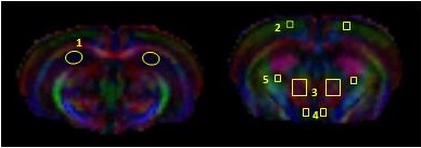
A total of 28 C57/BL6’ male mice (8 to 10 weeks old) were taken and randomly divided into four groups with 7 animals in each group. Mice were exposed to radiation dose of 5 Gy for both whole body (Group 1) and cranial (Group 2) through Tele 60Co irradiation facility. Controls (two groups) were sham irradiated. Brain MR Experiments were performed at 1 and 3 and 8 months post-irradiation(PI) on a 7T system (Bruker). DTI images were acquired using a multi-slice, multiple-shot spin echo EPI sequence with repetition time(TR)/echo time(TE) = 5000 ms/34.46 ms. The diffusion sensitive gradients were applied in 46 non co-linear directions at b=672 s/mm2. Java based DTI analysis software was used for the generation of FA(Fractional Anisotropy) and MD(Mean Diffusivity) maps.. Bilateral ROIs were manually drawn on Hippocampus(HIP), Sensory-motor cortex(SMC), Thalamus(TH), Hypothalamus(HY) and Caudoputamen(CP) regions as shown in Fig.1. FA and MD values from right and left hemisphere of these ROIs were considered together for statistical analysis. One-way analysis of variance (ANOVA) with multiple comparisons using Bonferroni, Post Hoc test was performed to evaluate the differences in DTI measures among different groups.Results
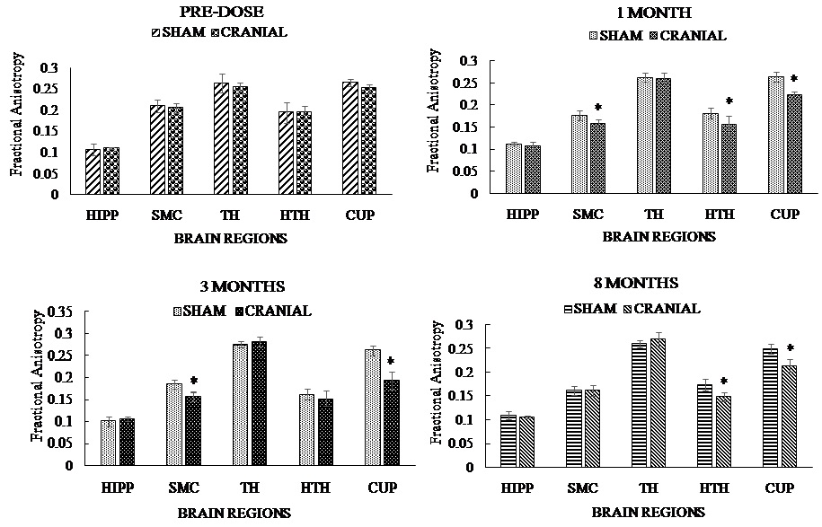
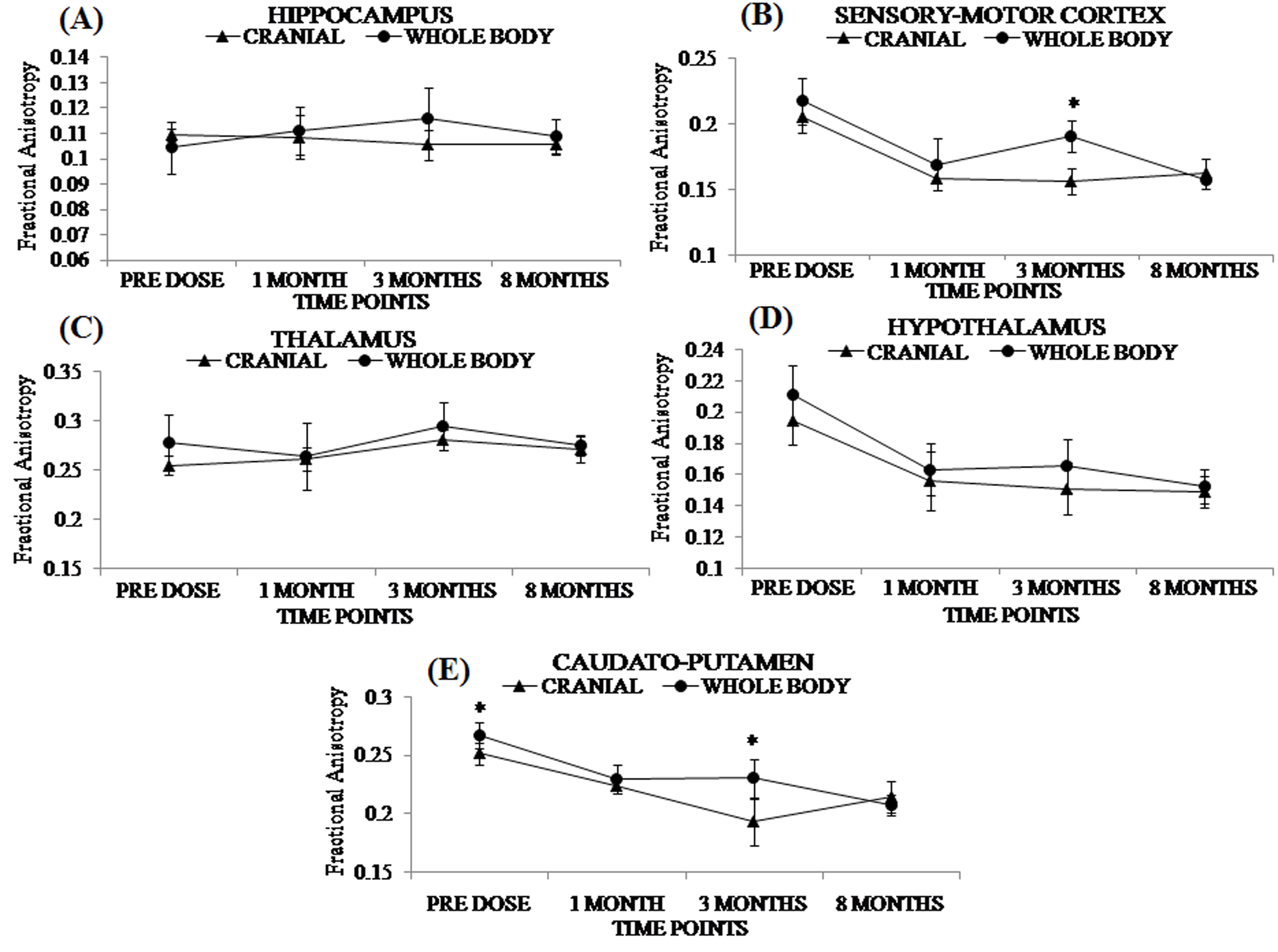
In our study, we observed a differential response of gray matter regions against whole body and cranial radiation. In cranial irradiation, FA values were significantly altered during sub acute phase in SMC, HY and CP at 1 month which continued to decrease till 8 months in CP. Whereas, maximum changes in MD was observed at early delayed phase i.e. 8 months PI wherein, most of the regions showed increased MD compared to controls. Contrary to cranial radiation induced FA changes, longitudinal DTI study did not show much FA changes in animals exposed to whole body radiation. Minimal or no change in FA was observed at all the time points except at 1 month. Interestingly, whole body radiation group showed decreased MD values in few regions in irradiated animals compared to controls till the end of the study which was strikingly in contrast to cranial irradiated groups that showed increased MD values compared to controls.Discussion
Nowadays use of DTI in gray matter region has emerged as an interesting area of research and a few recent DTI studies have reported increased diffusivities in gray matter in clinical pathologies4,5. Higher MD values are the characteristic of gray matter regions and increased MD values are mainly associated with increased extra cellular space and loss of tissue across the gray matter6. It is speculated that loss of glial progenitor cells due to radiation injury may be one of the reason for increased MD in different GM regions. Increased diffusivity in GM regions observed in our study are in accordance with some recent findings in pediatric patients where ADC values of these regions were found to be increased following cranial radiation therapy2,7. On the other hand, MD changes observed following whole body radiation exposure were just opposite of what it was observed after cranial radiation and that might be due to persistent neuro-inflammation occurring in hippocampus, in particular, following whole body radiation exposure.Conclusion
This study is a preliminary effort to characterize and compare sub-acute and early delayed micro structural changes in the gray matter regions of CNS after cranial and whole body radiation exposure using DTI. These preliminary findings would be useful to evaluate the mechanism of radiation pathophysiology and valuable in monitoring the treatment strategies and protocols during the course of radiation injury after either therapeutic or accidental radiation exposure.Acknowledgements
The present work is supported by Defence Research and Development Organisation (DRDO), Ministry of Defence, IndiaReferences
1. Gupta M, Rana P, Trivedi R et al. Comparative evaluation of brain neurometabolites and DTI indices following whole body and cranial irradiation: a magnetic resonance imaging and spectroscopy study. NMR Biomed 2013; 26:1733-1741
2. Horská A, Nidecker A, Intrapiromkul J et al. Diffusion tensor imaging of deep gray matter in children treated for brain malignancies. Childs Nerv Syst 2014; 30:631-638.
3. Nieman BJ, de Guzman AE, Gazdzinski LM et al. White and gray matter abnormalities after cranial radiation in children and mice. Int J Radiat Oncol Biol Phys 2015; 93:882-891.
4. Kumar M, Duda JT, Yoon SY et al. Diffusion Tensor Imaging for assessing brain gray and white matter abnormalities in a feline model of a Mannosidosis. J Neuropathol Exp Neurol 2015; 0:1-9.
5. Hannoun S, Durand-Dubief F, Confavreux C et al. Diffusion tensor-MRI evidence for extra-axonal neuronal degeneration in caudate and thalamic nuclei of patients with multiple sclerosis. Am J Neuroradiol 2012; 33:1363-1368.
6. Le Bihan D. Looking into the functional architecture of the brain with diffusion MRI. Nat Rev Neurosci 2003; 4:469-480.
7. Kim HJ, Kim SJ, Kim HS, et al. Alterations of mean diffusivity in brain white matter and deep gray matter in Parkinson's disease. Neurosci Lett 2013; 550:64-68.
Figures