5334
Eddy current characterization and correction using field monitoring for diffusion weighted imaging with maximal strength of 250 mT/m1Dept. of Radiology, Medical Physics, Medical Center University of Freiburg, Faculty of Medicine, Freiburg, Germany, 2Institute of Biomedical Engineering, National Taiwan University, Taipei, Taiwan, 3Athinoula A. Martinos Center for Biomedical Imaging, Department of Radiology, Harvard Medical School, Massachusetts General Hospital, Charlestown, MA, United States
Synopsis
We used a 24-channel field probe array to characterize the eddy current in a connectome scanner during
Introduction
Eddy current correction for diffusion-weighted imaging is important for elucidating high resolution features, because images with different diffusion sensitive gradients need to be registered to each other accurately to minimize blurring and signal loss1. Image distortion caused by eddy current is more serious when a stronger gradient is used2. The gradient amplitude in a connectome scanner is as high as 300mT/m3. This performance allows high b-value diffusion sensitive contrast to be established in a much shorter interval and consequently higher SNR. We hypothesize that eddy current artifact will be more serious in high b-value imaging in the connectome scanner. We attempted to characterize and to correct this artifact using field probes4.Method
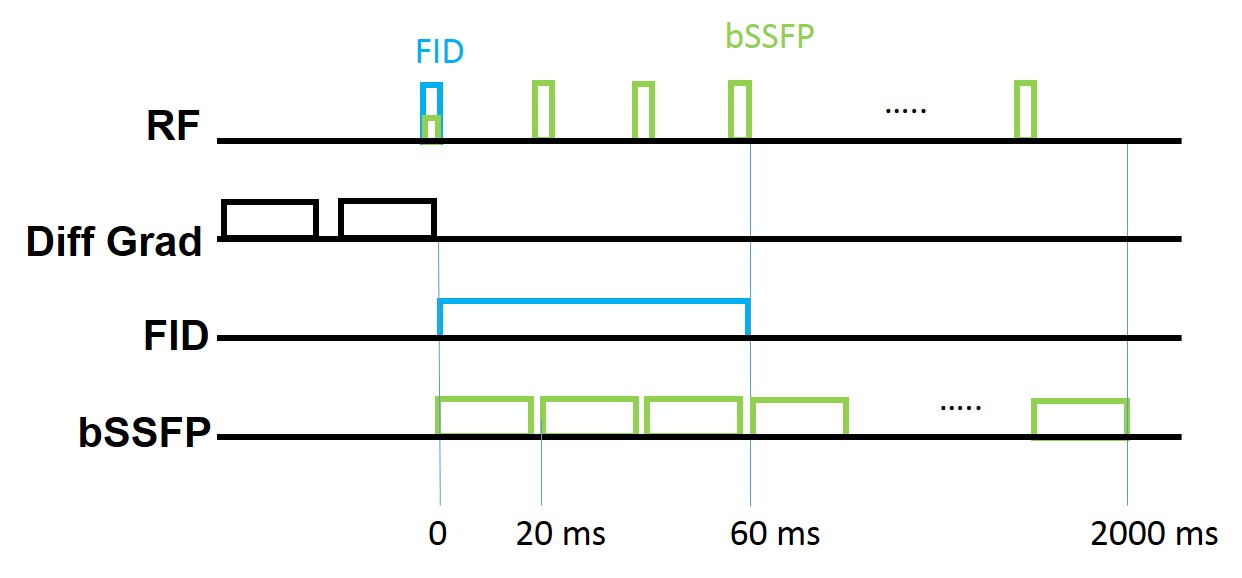
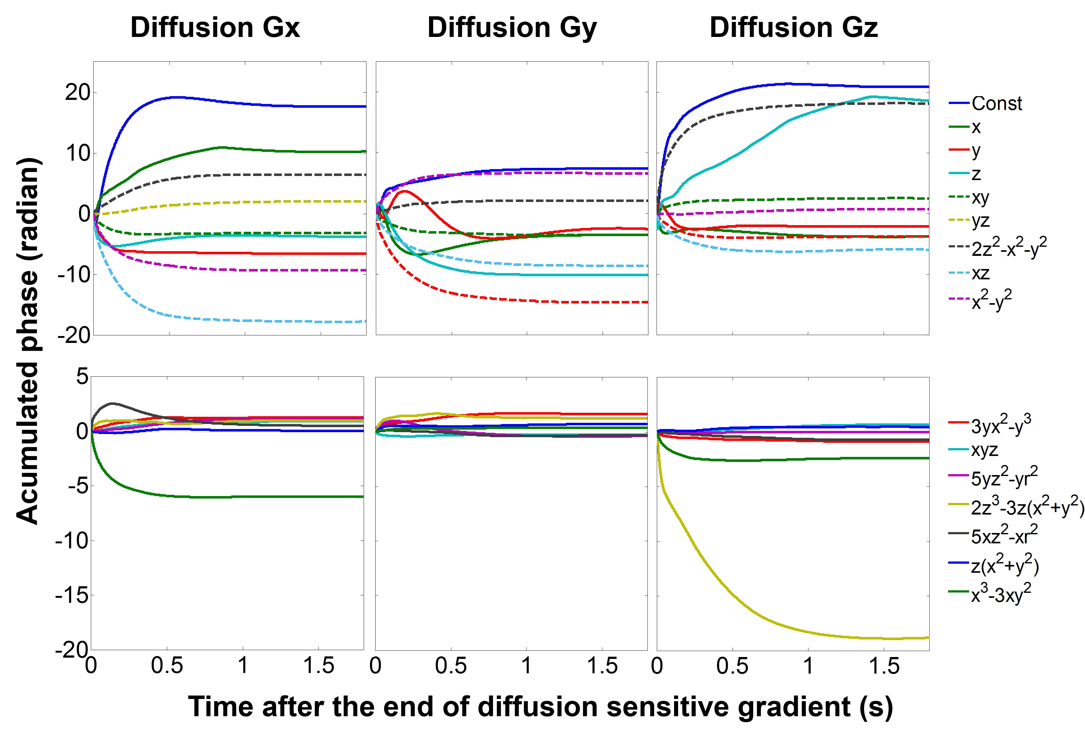
The magnetic field variation caused by diffusion sensitive gradient was measured using a 24-channel field probes array5. The magnetic field distribution up to the 3rd-order spherical harmonics was fitted from the probe measurements. The accumulated phase and strength for each spherical harmonic term was characterized by its maximum within a 10-cm diameter spherical phantom in order to compare the effects between different terms. We investigated the magnetic field generated by eddy current induced from the trapezoidal diffusion sensitive gradient waveforms with ramp time=2500 μs, flat top time=6950 μs and x-, y-, z-directional amplitude of +/-150 mT/m, +/-200 mT/m, and +/-250 mT/m. Because the eddy current may last longer than 1 s for each set of diffusion sensitive gradient, we used two pulse sequences with field probes to monitor the eddy current magnetic field separately. The first measurement used the FID sequence to characterize the eddy current right after diffusion sensitive gradients. As eddy current lasted much longer than the lifetime of probes (<100 ms), we used a bSSFP sequence (TR=20 ms) to monitor the slow-varying eddy current in the latter period in a separate measurement. The bSSFP sequence was repeated 3 times with central frequency at 0, 12 Hz, and 24 Hz to avoid signal void in bSSFP Details of the sequence were shown in Figure 1. A Stejskal-Tanner spin-echo diffusion weighted sequence was used in this study with imaging parameters: TR/TE=2600/83 ms, resolution=2 mm, b=5000 s/mm2. Note that x-, y-, and z-directional diffusion sensitive gradient used the same waveforms as those in field probe measurements. All measurement were done on the connectome scanner (Siemens).Results
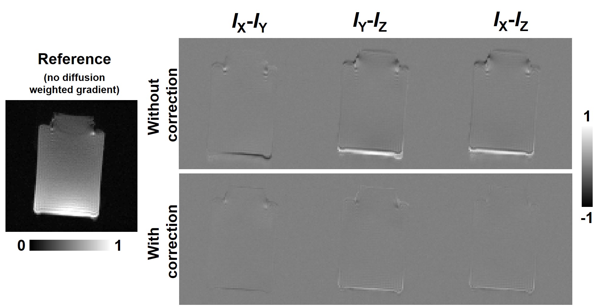
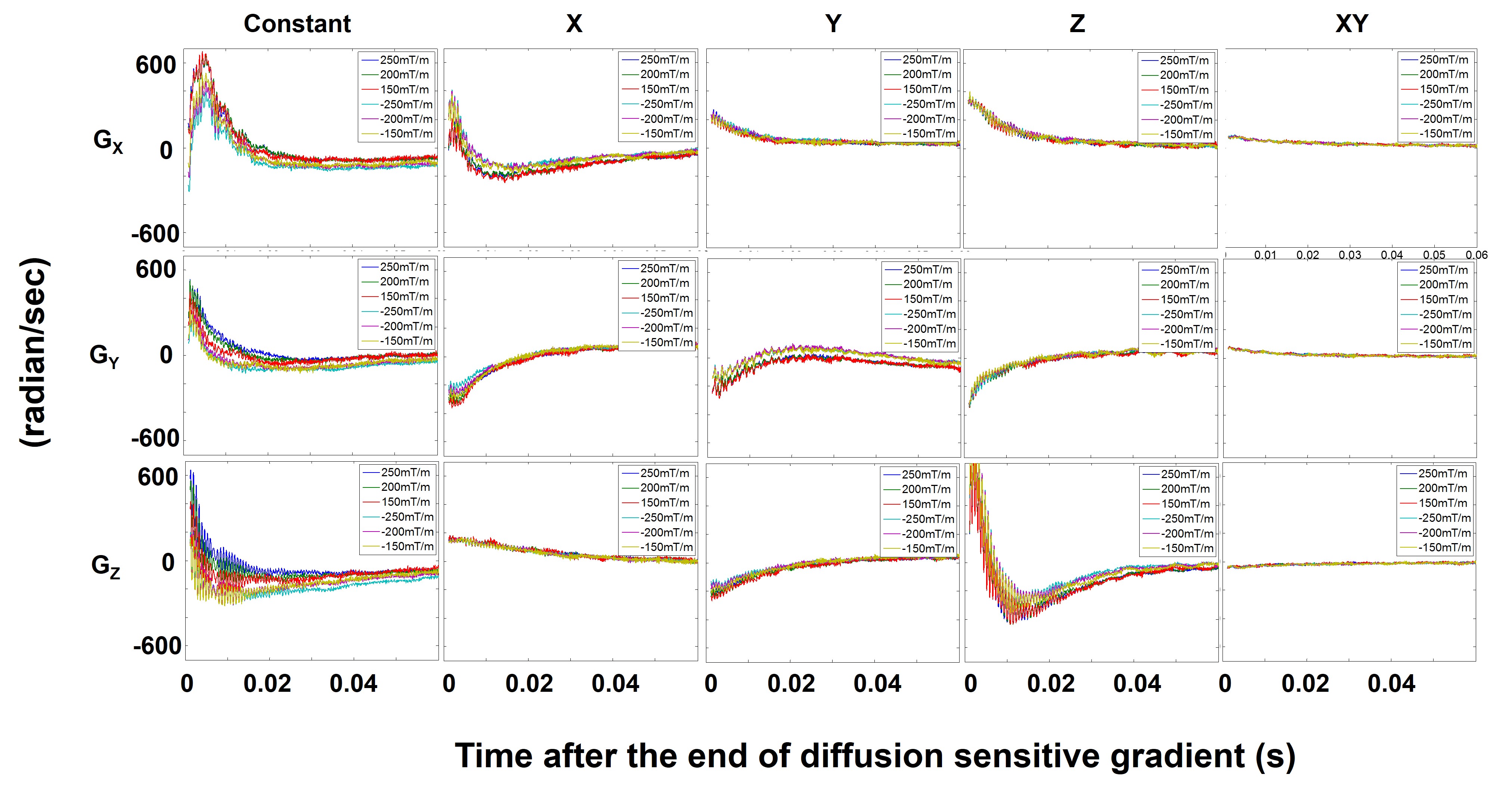
Figure 2 shows the measured accumulated phase caused by eddy current of x, y, and z diffusion sensitive gradient with 250 mT/m amplitude. The normalized accumulated phase of the linear terms of the fitted spherical harmonic still showed prominent changes after 1 s by the end of diffusion sensitive gradient, suggesting the long lifetime of eddy current. Strong and long-lasting (>1 s) eddy current was also observed on 2nd and 3rd-order terms. Figure 3a shows the diffusion-weighted images with bx=by=bz=5000 s/mm2 and their differences without eddy current correction. Figure 3b shows images after eddy current correction. The difference between images with diffusion sensitive gradient in different directions became smaller, suggesting reduced distortion caused by different eddy currents. Figure 4 shows the scaled strength for the fitted spherical harmonics as x-, y- and z-direction diffusion sensitive gradient at 6 different strengths. The scaling was chosen to be the ratio between the maximum gradient strength (250 mT/m) and the used gradient strength. The waveforms of constant and linear field depended on the gradient amplitude and polarity. Non-overlapping among scaled strength curves suggested that the gradient system was not a linear time-invariant system. Figure 4 only shows the xy nonlinear term, because other nonlinear terms behaved similarly by showing overlapped scaled strength curves.Discussion
Combining FID and bSSFP sequences, we monitored the magnetic field change due to eddy current lasting longer than 1 s on the connectome scanner, suggesting the necessary duration to monitor the magnetization history in order to characterize eddy current artifacts accurately. The difference between diffusion-weighted images was reduced after correction but still visible (Figure 3). Using higher than the 3rd-order spherical harmonics may achieve better correction by improving the eddy current characterization. Our results suggested that the gradient system in the connectome scanner is not a linear time-invariant system (Figure 4). Thus, the eddy current artifact in images with a specific combination of diffusion sensitive directions may not be successfully predicted as the sum of image artifacts with diffusion sensitive gradient acting alone. Therefore, a field monitoring system that allows detecting the eddy current and imaging data concurrently is important6.Acknowledgements
This work was partially supported by Ministry of Science and Technology, Taiwan (103-2628-B-002-002-MY3, 105-2221-E-002-104, 106-2911-I-002-502), and the Academy of Finland (No. 298131).
References
1. Tuch, D. S. et al. High angular resolution diffusion imaging reveals intravoxel white matter fiber heterogeneity. Magnetic resonance in medicine 48, 577-582 (2002).
2. Mangin, J.-F., Poupon, C., Clark, C., Le Bihan, D. & Bloch, I. Distortion correction and robust tensor estimation for MR diffusion imaging. Medical image analysis 6, 191-198 (2002).
3. Sotiropoulos, S. N. et al. Advances in diffusion MRI acquisition and processing in the Human Connectome Project. Neuroimage 80, 125-143 (2013).
4. Barmet, C., Zanche, N. D. & Pruessmann, K. P. Spatiotemporal magnetic field monitoring for MR. Magnetic Resonance in Medicine 60, 187-197 (2008).
5. Chu, Y.-H., Hsu, Y.-C. & Lin, F.-H. Decoupled dynamic magnetic field measurements improves diffusion-weighted magnetic resonance images. Scientific Reports 7, 11630 (2017).
6. Barmet, C. et al. in Proceedings of the 18th Annual Meeting of ISMRM, Stockholm, Sweden. 216.
Figures