5333
SPIRiT-Based Reconstruction of Interleaved Multi-shot EPI (SPECTRA) for Navigator-Free Diffusion Weighted MRI1Advanced Application, Alltech Medical Systems, Chengdu, China, 2Alltech Medical Systems, Chengdu, China
Synopsis
Multi-shot EPI can achieve a high imaging resolution for Diffusion weighted MRI (DWI) by sampling a large k-space without increasing the degree of T2* blurring. However, its shot-to-shot phase error has to be corrected. Recently, a navigator-free correction method based on SENSE technique has been developed, assuming the phase error is spatially smooth among different DWI shots. In this work, we propose a SPIRiT-based method to correct the phase error and iteratively reconstruct navigator-free multi-shot EPI. Our results suggest it has the advantage of insensitivity to coil map bias and better SNR compared to the SENSE-based method.
Purpose
A SPIRiT-based reconstruction algorithm is proposed to iteratively generate diffusion-weighted images (DWI) acquired by navigator-free interleaved multi-shot EPI.Introduction
Diffusion-weighted magnetic resonance imaging (DWI) has become more and more popular as a clinical diagnosis tool [1-4]. Conventional DWI data are usually acquired with single-shot echo-planar imaging (EPI) due to its high scan efficiency and motion immunity [5-7]. However, single-shot DWI is fundamentally constrained by available acquisition time due to T2* decay. Hence only limited image spatial resolution can be achieved even when parallel imaging is used [8-9]. This makes it difficult to detect subtleties in fine structures where a high spatial resolution is required. This problem can be largely alleviated using a multi-shot acquisition. However, multi-shot DWI is very sensitive to shot-to-shot motion-induced phase errors due to the diffusion sensitizing gradients, leading to signal cancellation or aliasing artifacts [10]. Recently, a SENSE-based reconstruction has been proposed to efficiently correct the shot-to-shot phase errors from navigator-free multi-show DWI, assuming the phase error is spatially smooth among different DWI segments [11-12]. One challenging aspect of it is that its image reconstruction quality highly depends on the quality of the estimated coil sensitivity maps, especially at the regions with low SNR. In this work, we propose a SPIRiT-based reconstruction algorithm (SPECTRA), which does not use coil sensitivity maps, to correct the phase error and iteratively reconstruct navigator-free DWI images acquired by interleaved multi-shot EPI.Theory & Method
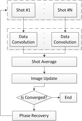
A DWI (1.0 X 1.0 X 4.0 mm3) dataset was acquired with a 3-shot interleaved EPI sequence from a healthy volunteer on a 1.5T MRI system (Alltech Centauri, Chengdu, China) equipped with an 8-channel head coil. The dataset consisted of one baseline image with a b-value =0 and three images with a b-value = 1000 s/mm2. Diffusion gradients were applied along three orthogonal directions and averaged three times in each direction. In-plane acquisition matrix was 512 X 222. Partial Fourier factor was set to 0.8 along the phase encoding direction. A single-shot EPI with a b=0 was used as the pre-scan for SPIRiT calibration and coil sensitivity estimation. The acquired data were processed as shown in the flow chart in Fig. 1. First, a 5X5 SPIRiT kernel was calculated from the pre-scan data. Second, data convolution was performed with SPIRiT kernel for each DWI shots. Third, a Hanning window smoothed phase map Vk was generated for each shot using Eqn [1]. If multiplying its conjugate Vk* with Ik, only the high-frequency phase variations of Ik remain, with the magnitude of Ik unchanged. Then a shot-combined, smooth phase variation filtered DWI image Iavg can be generated after a normalization using Eqn [2]. The underlying assumption is that the phase error across different shots are spatially smooth and the images from different shots have the same magnitude. After that, individual shot images were updated using Eqn [3] and went on to the next iteration until stop criteria was met. Finally, a 10-iteration POCS-based Partial Fourier reconstruction was performed before the sum-of-square channel combination.
$$V_{k} = \frac{Hann(I_{k})}{|Hann(I_{k})|}\qquad\qquad\qquad\qquad\qquad\qquad\quad[1]$$
where $$$I_{k}$$$ is the image from $$$K_{th}$$$ shot
$$I_{avg} = \sum_k^N(W_{k}I_{k}), where \quad W_{k} = \frac{V_k^*}{\sum_k^N(|V_{k}|)}\qquad[2]$$
$$I_k^{(n+1)} = I_{avg}^{(n)}V_k^{(n)}\qquad\qquad\qquad\qquad\qquad\qquad\quad[3]$$
Results
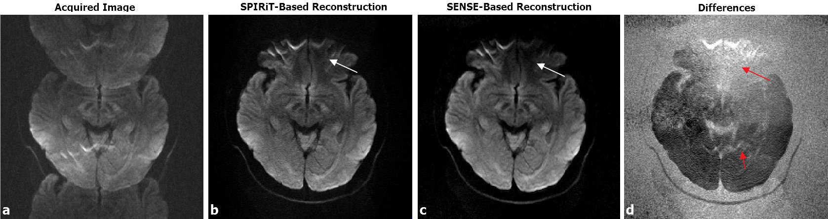
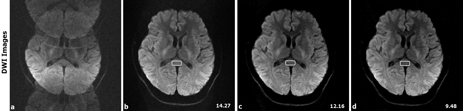
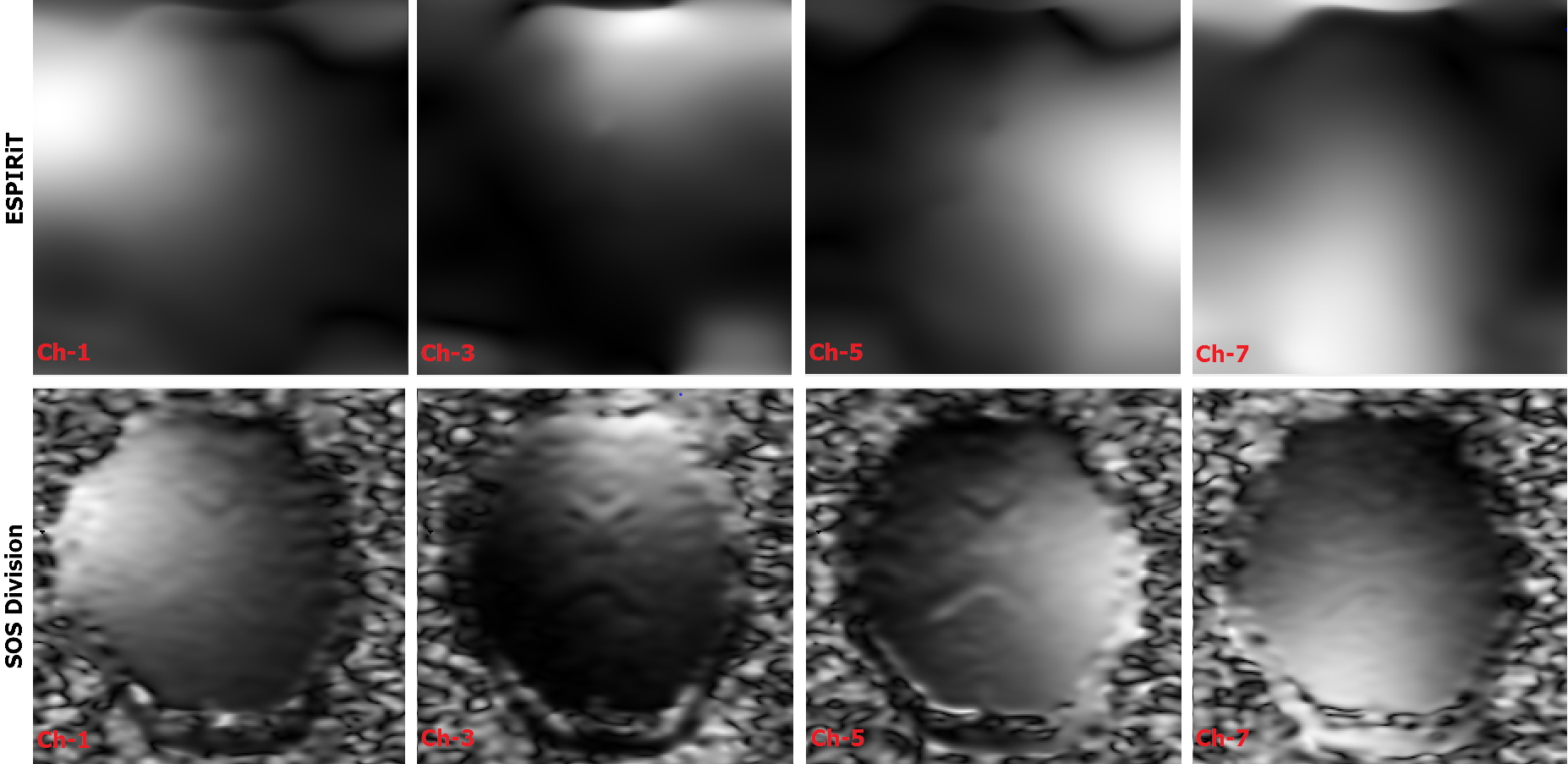
Figure 2 demonstrates images reconstructed using the proposed and the previous SENSE-based method, respectively. The SPIRiT-based method shows less signal cancellation at regions with low SNR, as indicated by the white arrows in the images (b) and (c). The SENSE reconstruction has a larger signal cancellation due to its incomplete unfolding of aliased images, which is quite obvious on the difference image (d). To compare image SNR, we measured the ratio of the mean value to the standard deviation within a region of interest (ROI) placed in the center of each image (Figure 3). The SNR of SPIRiT-based method was 14.27, while that of SENSE-based method was 12.16 (if estimating sensitivity map using ESPIRiT) and 9.48 (if estimating sensitivity map using SOS division),respectively.Figure 4 presents coil sensitivity maps estimated with ESPIRiT algorithm and SOS-division method, respectively.Discussion and Conclusion
In this study, we present a SPIRiT-based method to correct the shot-to-shot phase error in navigator-free multi-shot DWI. Compared to the traditional SENSE-based method, the proposed method is less sensitive to the bias from the coil sensitivity map estimation, hence providing images with a higher SNR. The reconstruction quality can be further improved by incorporating additional prior knowledge such as the low-rank constraint and global sparsity, which will be our future work.Acknowledgements
No acknowledgement found.References
[1] Le Bihan D, Breton E, Lallemand D, Aubin ML, Vignaud J, Laval-Jeantet M. Separation of diffusion and perfusion in intravoxel incoherent motion MR imaging. Radiology. 168, 497–505(1988).[2] Moseley ME, Cohen Y, Kucharczyk J, Mintorovitch J, Asgari HS, Wendland MF, Tsuruda J, Norman D. Diffusion-weighted MR imaging of anisotropic water diffusion in cat central nervous system. Radiology. 176, 439–45(1990).[3] Basser PJ, Mattiello J, LeBihan D. MR diffusion tensor spectroscopy and imaging. Biophys J. 66, 259–267 (1994).[4] Muller MF, Prasad PV, Bimmler D, Kaiser A, Edelman RR. Functional imaging of the kidney by means of measurement of the apparent diffusion coefficient. Radiology. 193, 711–715 (1994).[5] Jezzard P, Balaban RS. Correction for geometric distortion in echo planar images from B0 field variations. Magn Reson Med 34, 65–73(1995).[6] Turner R, Le Bihan D, Chesnick AS. Echo-planar imaging of diffusion and perfusion. Magn Reson Med. 19, 247–253 (1991).[7] Anderson AW, Gore JC. Analysis and correction of motion artifacts in diffusion weighted imaging. Magn Reson Med. 32, 379–387 (1994).[8] Pruessmann KP, Weiger M, Scheidegger MB, Boesiger P, SENSE: Sensitivity Encoding for Fast MRI. Magn. Reson. Med. 42, 952–962 (1999).[9] Griswold MA, Jakob PM, Heidemann RM, Mathias Nittka, Jellus V,Wang J, Kiefer B, Haase A. Generalized autocalibrating partially parallel acquisitions (GRAPPA). Magn. Reson. Med. 47, 1202-1210 (2002).[10] Butts K, de Crespigny A, Pauly JM, Moseley M. Diffusion-weighted interleaved echo-planar imaging with a pair of orthogonal navigator echoes. Magn Reson Med 35, 763–770 (1996).[11] Chen NK, Guidon A, Chang HC, Song AW. A robust multi-shot scan strategy for high-resolution diffusion weighted MRI enabled by multiplexed sensitivity-encoding (MUSE). Neuroimage 72, 41–47 (2013).[12] Guo H, Ma X, Zhang Z, Zhang B, Yuan C, Huang F. POCS-enhanced inherent correction of motion-induced phase errors (POCS-ICE) for high resolution multishot diffusion MRI. Magn ResonMed 75, 169–180 (2016).Figures