5331
Randomized-Slice Simultaneous Multi-Slice for Diffusion Kurtosis Imaging1Biophysics, Medical College of Wisconsin, Milwaukee, WI, United States, 2Radiology, Medical College of Wisconsin, Milwaukee, WI, United States, 3Neurosurgery, Medical College of Wisconsin, Milwaukee, WI, United States
Synopsis
Simultaneous Multi-slice (SMS) techniques excite multiple slices simultaneously to accelerate MRI data acquisition. However, slice separation during image reconstruction is not exact and results in coupling between separated voxels. While this may not be critical for most anatomic imaging methods, small but consistent leakage of information from another slice in a DKI dataset will cause bias in diffusion parameter estimates. Here, we implement a randomized-slice pairing technique to alleviate this problem in diffusion MRI acquisitions.
Introduction
Simultaneous Multi-slice (SMS) techniques reduce acquisition times by exciting multiple slices simultaneously and separating the aliased slices using a mathematical model. However, this slice separation is not exact and leads to information leakage between simultaneously excited slices.1,2 Thus, measurements in a voxel will be affected by the residues from the overlapped slices. In diffusion MRI techniques, such as DTI or diffusion kurtosis imaging (DKI), this introduces a significant problem. If the same group of slices are excited simultaneously across all diffusion weighted images, the same voxels are mixed every time, and the diffusion estimates in each voxel are biased by the slice leakage. Earlier, we proposed a novel adaptation of SMS to mitigate the effects of slice leakage in diffusion metrics. We randomize which slices are simultaneously excited across different diffusion weighted images. Since overlapping slices will be different for each diffusion weighted image, residual effects from imperfect slice separation will appear as random noise. With enough diffusion directions and weightings, the model parameter estimation should be robust against this random noise. We refer to this method as randomized-slice (rs-)SMS. The simulation results that we presented last year demonstrated that the accuracy of DKI metrics estimated from rs-SMS was higher compared to standard SMS. However, those simulations did not take into account various technical limitations that needed to be considered in actual implementation. We recently implemented the rs-SMS pulse sequence and image reconstruction software and conducted in vivo experiments. Here we present results from DKI scans acquired with conventional single-band, conventional SMS and rs-SMS.Methods
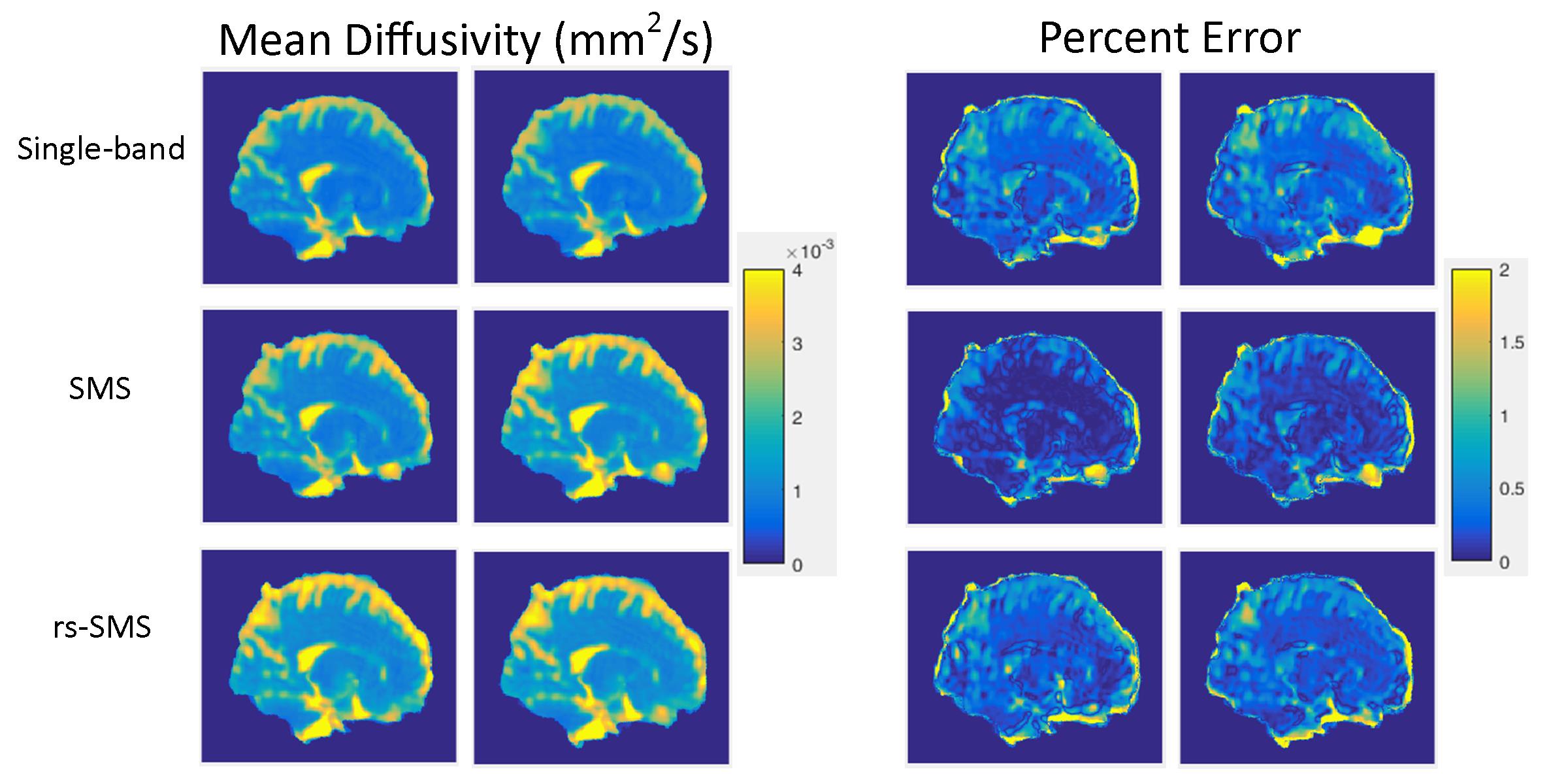
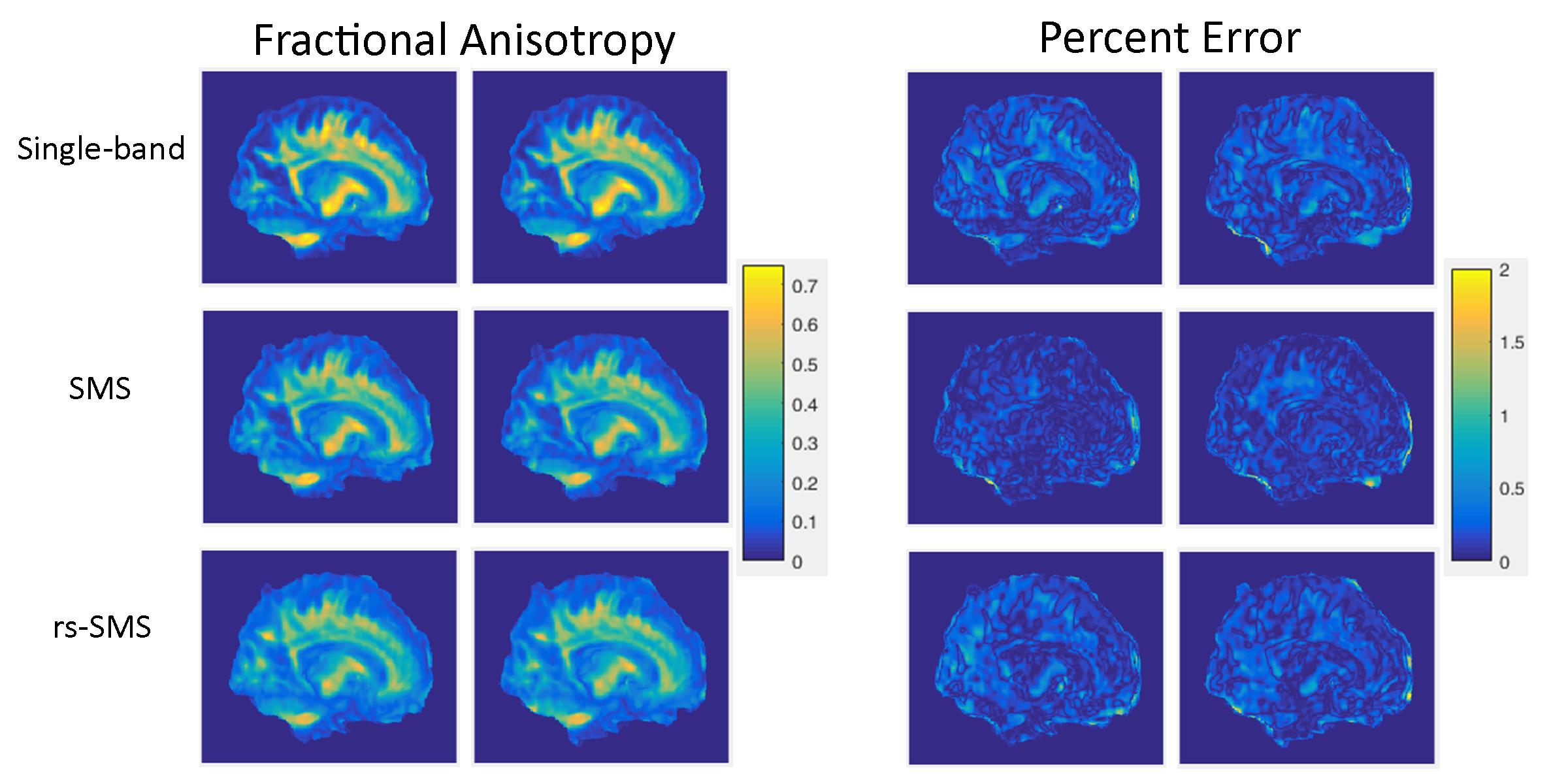
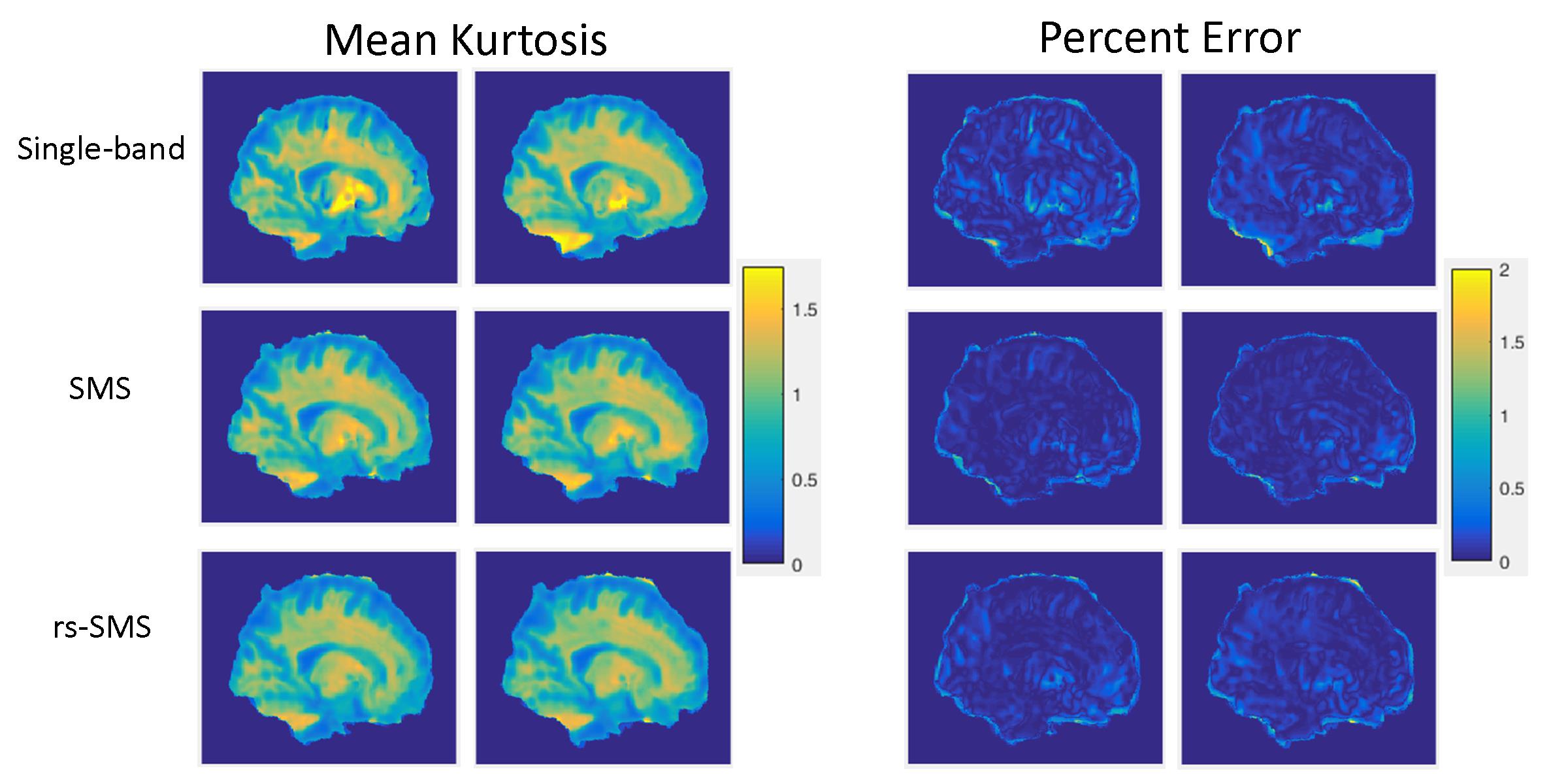
Data were acquired from a healthy subject (M,35yrs). The study was approved by the IRB and written consent was obtained from the subject. Diffusion data were acquired with a GE MR950 7T MRI scanner and single-shot SE-EPI sequence with 3mm isotropic voxels, one non-diffusion weighted image and diffusion-weighted images with b=1000s/mm2 and 2000s/mm2 (30 diffusion directions for each shell). TE/TR: 97.3/3250ms, and the acquisition matrix was 80x80x48. Two scans each for single-band, SMS, and rs-SMS were acquired for a total of 6 acquisitions. Susceptibility distortion correction was performed with TOPUP and eddy current distortion correction was performed with eddy of FSL.3 Diffusion metrics of mean diffusivity (MD), fractional anisotropy (FA), and mean kurtosis (MK) were calculated. Images were registered to a standard template for comparisons using ANTs diffeomorphic registration software.4Results
A representative slice for the three metrics MD (figure1), FA (figure 2), and MK (figure 3) is presented for each acquisition: single-band (Row 1), SMS (Row 2), and rs-SMS (Row3). Columns 1 and 2 are the metric maps for trial 1 and 2, respectively. Columns 3 and 4 are the percent error maps relative to the average from all six scans. All percent error maps are windowed at 0-2% error, regardless of the diffusion metric.Discussion
Qualitatively, DKI metric images were comparable in both SMS and rs-SMS to single-band. The percent error maps illustrate diffuse differences of around 1% error for all metrics. Outlier voxels on the image perimeter are attributed to misregistration of the images. Variability between single-band and both multiband techniques is within the interscan differences for single-band. With more subjects, a statistical analysis could quantitatively determine the effect of multiband diffusion MRI on DKI metrics.
Technical challenges were predominantly slice cross-talk and minimum slice distance restraints. Minimum slice distance for multiband excitation was required to make use of coil sensitivity differences for effective slice separation in reconstruction. So, the minimum slice separation was set to 35mm for simultaneously-excited slices to leverage coil sensitivity differences in slice separation. However, this limited the number of possible slice combination patterns of rs-SMS. Slice crosstalk due to imperfect slice profiles and partial excitation of spins in neighboring slice are usually minimized by interleaved slice acquisition scheme. However, in rs-SMS one needs to pay close attention to avoid exciting adjacent slices shortly after one another. Careful SMS randomization scheme to maximize the time between excitations of adjacent slices, and better characterization of the slice profiles generated by the multiband RF pulses are essential to minimize such artifacts. These design criteria help reduce such slice cross-talk effects that lead to signal attenuation in the diffusion-weighted images from neighboring slices. Another consideration was to limit repeated slice combinations in the diffusion weighted image set. Due to limited number of slices acquired and constraints on minimal slice distance for simultaneous excitation some slice pairs had to be repeated for 60-direction diffusion acquisition. With the given constraints, one needs to find optimal slice combination patterns for rs-SMS.
Acknowledgements
No acknowledgement found.References
1. Cauley SF et al. (2014) Interslice leakage artifact reduction technique for simultaneous multislice acquisitions.Magnetic Resonance in Medicine 72:93-102.
2. Setsompop K et al. (2013) Pushing the limits of in vivo diffusion MRI for the Human Connectome Project. NeuroImage 80:220-233.
3. Jenkinson M et al. (2012) FSL. NeuroImage, 62:782-90
4. Avants BB et al. (2008) Symmetric diffeomorphic image registration wtih cross-correlation: Evaluating automated labeling of elderly and neurodegenerative brain. Medical Imaging Analysis 12:26-41.
Figures