5328
Combined Crushers: Omitting Separate Crusher Gradients in the Twice-Refocused Spin Echo Diffusion MRI Sequence1Institute of Neuroscience and Medicine - 4, Jülich Research Centre, Juelich, Germany, 2RWTH Aachen University, Faculty of Medicine, Department of Neurology, JARA, Aachen, Germany
Synopsis
A new method is proposed that allows for the omission of crusher gradients in the Twice-Refocused Spin Echo (TRSE) sequence. The proper spoiling of the unwanted signal pathways is achieved by modifying the amplitudes and durations of the diffusion gradients. Both the crusher moment as well as the desired diffusion weighting is eventually provided only by the diffusion gradients. This results in several advantages like shorter minimum TEs and higher SNRs, all of which can be beneficial in diffusion MRI.
Introduction
The Twice-Refocused Spin Echo (TRSE) diffusion sequence by Reese et al.1 is normally executed with four separate crusher gradients alongside the two refocusing RF pulses (see Fig. 1 top). The crusher gradients are necessary to spoil unwanted signal pathways while maintaining the desired TRSE pathway. There are several possibilities regarding the orientation of the crusher gradients, e.g. orthogonal to the diffusion gradients2. It is impractical to apply separate crushers (anti-)parallel to the diffusion gradients since this could lead to cancellation effects compromising the crusher efficiency2. However, having four separate crusher gradients not aligned with the diffusion gradients is time-consuming and leads to a TE that is longer than inherently necessary in diffusion MRI as well as a correspondingly lower SNR. Besides, the common design introduces a bias in the b-value due to the crusher contributions off the principal diffusion direction.
Here, we propose a new crusher concept which circumvents both drawbacks. Our approach is to omit separate crusher gradients completely and, instead, modify the diffusion gradient amplitudes and durations (see Fig. 1 bottom) such that both correct diffusion weighting and proper crushing is provided only by the diffusion gradients. Using the combined crusher scheme it is possible to either reduce the minimum TE, decreasing the total scan time and increasing the SNR, or to increase the maximum possible b-value or combinations thereof.
Methods
The combined crusher method was implemented in an in-house TRSE diffusion EPI sequence. A calculation method was developed to determine the crusher moments, Ci, which have to be added to the diffusion gradients (see Fig. 1 bottom) to reliably spoil all seven unwanted signal pathways while preserving the desired TRSE signal. Note that depending on the timing and the b-value, the diffusion gradients might already provide enough crusher moment themselves in which case the Ci can be zero. In either case, care was taken such that the modified diffusion gradients (with moment Di+Ci, see Fig. 1 bottom) provide the desired b-value without bias, i.e. any diffusion gradient modifications are taken into account for the b-value calculation.
To prove the effectiveness of our method, phantom and in vivo experiments were performed with conventional and combined crushers on a commercial 3T Trio MRI scanner (Siemens Healthineers, Erlangen, Germany). The body coil was used for excitation, a 12-element phased array head coil was used for signal reception. Prior to the in vivo measurements written informed consent was obtained from a single healthy male volunteer.
The following sequence parameters were used in all experiments: TR=2000ms, resolution 1.8×1.8×3.0 mm3, matrix size 128×128, no. of slices 10, distance factor 100%. Six diffusion directions, comprising the positive and negative directions along the three gradient axes, were measured. The experiments were performed with the standard TE, which is possible with conventional and combined crushers, and a reduced TE which is only possible with combined crushers.
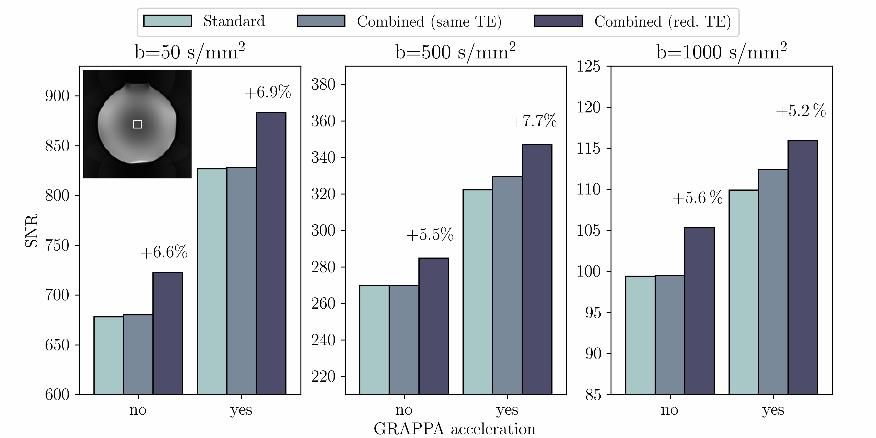
From the in vivo images, apparent diffusion coefficient (ADC) maps were computed after reconstruction and visually inspected for artefacts. A spherical water phantom was used to assess the expected SNR gains. The mean SNR was computed in a ROI by repeating the measurement in one diffusion direction 100 times and dividing the mean signal magnitude in every voxel by the corresponding standard deviation. Raw data reconstruction was performed with the online reconstruction system of the scanner. Post-processing for SNR and ADC calculations was performed using MATLAB (The Mathworks Inc., Natick, USA).
Results
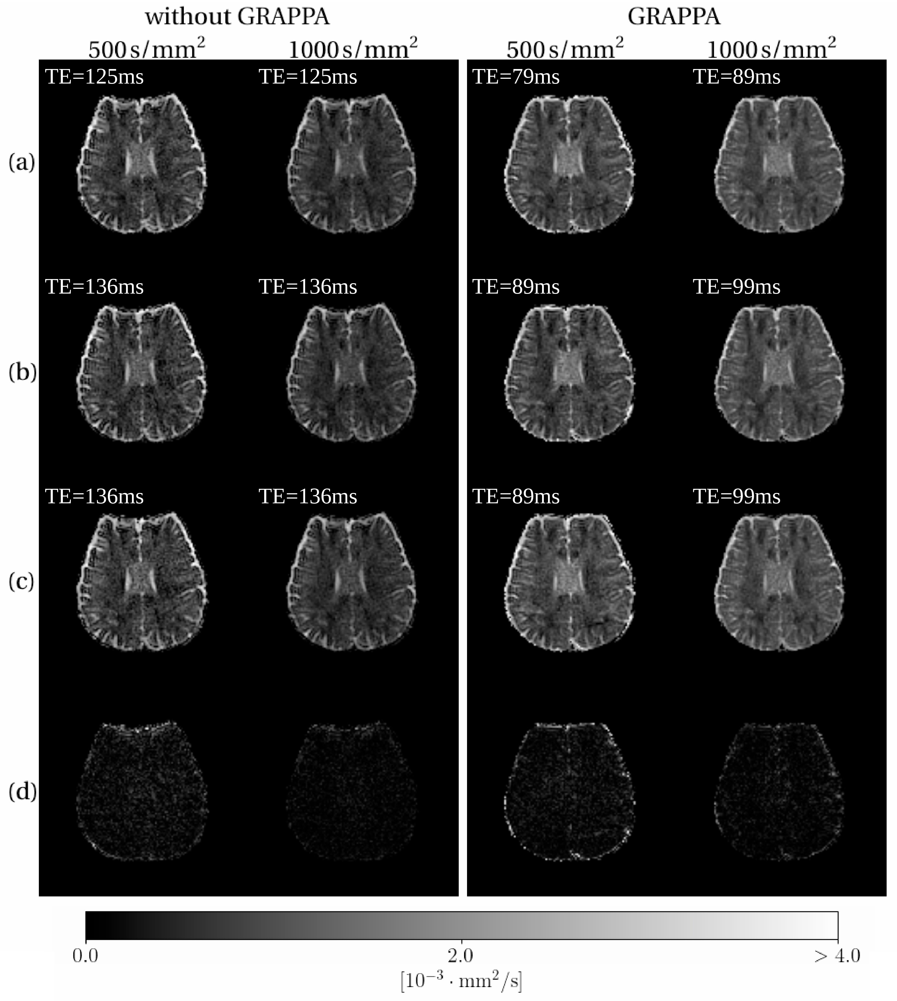
Figure 2 shows the ADC maps acquired with both sequences at the two different TE. Based on visual inspection, the modified sequence provides artefact-free ADC maps while the echo time could be reduced by 10 ms and 11 ms depending on the b-value and used GRAPPA factors. Figure 3 shows the result of the phantom measurement to assess the SNR gains. The SNR in the ROI was increased by 5.2% to 7.7%.Discussion and Conclusions
Our experiments demonstrate that the combined crusher concept is feasible and visual inspection of the ADC maps shows that it can provide artefact-free DWI results comparable to conventional crusher schemes. However, the proposed method allows one to improve several aspects of the TRSE diffusion sequence whereby the most prominent are a reduction of the minimum possible TE and an SNR increase. Although the original TRSE design1, aiming at optimal eddy-current suppression, is slightly changed, no eddy current effects have arisen during our investigations. In fact, numerical simulations suggest that omitting the separate crusher gradients (which were not considered for eddy current suppression in the original design) can even be beneficial in terms of reducing the overall eddy currents.
All in all, the combined crusher approach could become helpful for improving the performance and feasibility of diffusion MRI.
Acknowledgements
No acknowledgement found.References
1Reese T, Heid O, Weisskoff R, Wedeen V. Reduction of Eddy-Current-Induced Distortion in Diffusion MRI Using a Twice-Refocused Spin Echo. Magnetic Resonance in Medicine 2003;49:177–182
2Nagy Z, Thomas DL, Weiskopf N. Orthogonalizing crusher and diffusion-encoding gradients to suppress undesired echo pathways in the twice-refocused spin echo diffusion sequence. Magnetic Resonance in Medicine 2014;71:506–515
Figures
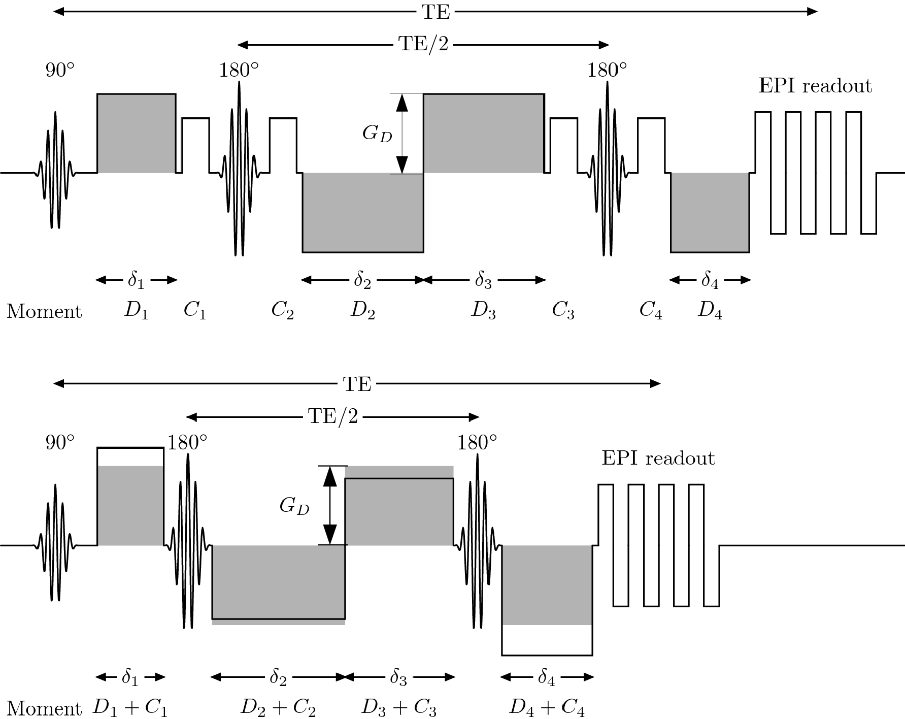
Figure 1: Schematic comparison of the conventional TRSE sequence with separate crusher gradients (top) and the combined crusher version without (bottom).
The original diffusion gradient amplitude, GD, and the gradient durations δi are modified in the combined crusher scheme. The diffusion and crusher gradient moments Di and Ci are calculated to assure proper spoiling of all unwanted pathways while simultaneously providing the desired diffusion weighting in the correct direction. Note that at maximum amplitude, the Di provide enough crusher moment themselves such that all Ci can be zero and the maximum allowed gradient amplitude is not exceeded.