5322
Distortion-Free High-Resolution Diffusion MRI with RPG-MUSE1Brain Imaging and Analysis Center, Duke University, Durham, NC, United States
Synopsis
Diffusion-weighted images acquired with multi-shot EPI at high-resolution are susceptible to inter-shot motion artifacts and geometric distortions resulting from magnetic-field inhomogeneities. This study presents an efficient means of inherently accounting for both motion and distortion artifacts by alternating the phase encoding gradient polarities in odd/even shots of multi-shot EPI. When acquired in this fashion, the proposed RPG-MUSE model simultaneously accounts for both shot-to-shot motion induced artifacts and geometric distortions during the reconstruction of multi-shot diffusion weighted images. This technique requires no additional scan time, and accounts for B0 and eddy current induced distortions specific to each diffusion-weighted volume.
Purpose
Recent advances in multi-shot echo-planar-imaging (ms-EPI) methodologies have seen increased utility in achieving high-resolution diffusion MRI (dMRI). Due to the prolonged readout time associated with sizeable acquisition matrices, however, the spatial distortions resulting from B0 and eddy current induced magnetic field (B-field) inhomogeneities remain a challenge in dMRI acquisitions. To address these challenges, this study presents an efficient and effective method of inherently accounting for both B-field distortions and shot-to-shot motion induced artifacts common in multi-shot dMRI. This is achieved by incorporating reversed polarity gradients (RPG)1,2,3 along the phase encoding direction (PE) into a multiplexed sensitivity encoding (MUSE)4 acquisition. Specifically, this technique, termed RPG-MUSE, applies opposite PE gradient polarities to odd/even shots of the ms-EPI acquisition scheme employed in a conventional MUSE scan, thereby allowing B0 and eddy currents induced distortions to be estimated for each diffusion volume individually, without increasing the acquisition time.Methods
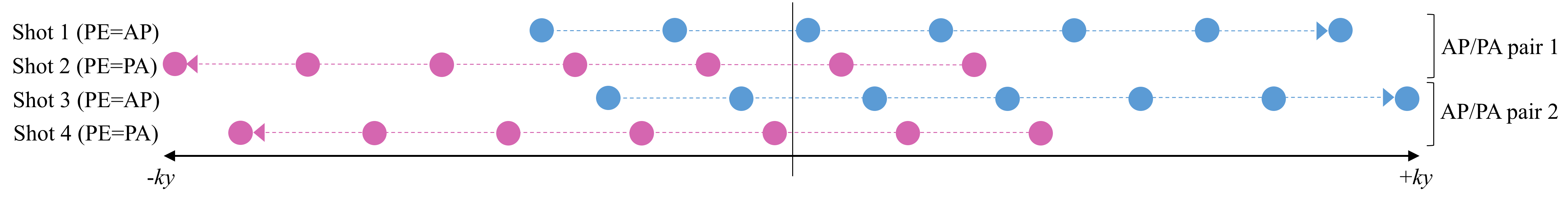
Whole brain in-vivo multi-shot DWI data was acquired in a commercial 3T GE MR750 scanner using a 32-channel head coil. Volumes with b=0 and b=800s/mm2 were acquired with 1.2x1.2x1.2mm3 isotropic voxels in 80 axial slices with a 30.7cm FOV and a 256x256 acquisition matrix. Four interleaved shots were acquired with PE=Anterior/Posterior in all odd shots and PE=Posterior/Anterior in all even shots (66% partial-Fourier along PE), as illustrated in Fig. 1. The interleaved shots were individually reconstructed with SENSE5 to produce four full-FOV shot images (two AP/PA pairs) for each slice in each dMRI volume. These shot images were then used to estimate 1) low frequency motion-induced phase variations between shots, and 2) B0/eddy current induced distortion maps for each AP/PA shot pair using FSL’s topup tool2,3. The shot phase images and field distortion maps were then fed into RPG-MUSE to reconstruct the raw interleaved ms-EPI data into full FOV slice images while simultaneously accounting for shot-to-shot motion artifacts and B-field distortions. Finally, a bias-field correction was applied to volumes generated from shot 1 (PE=AP), shot 2 (PE=PA), and RPG-MUSE using FSL6.Results and Discussion
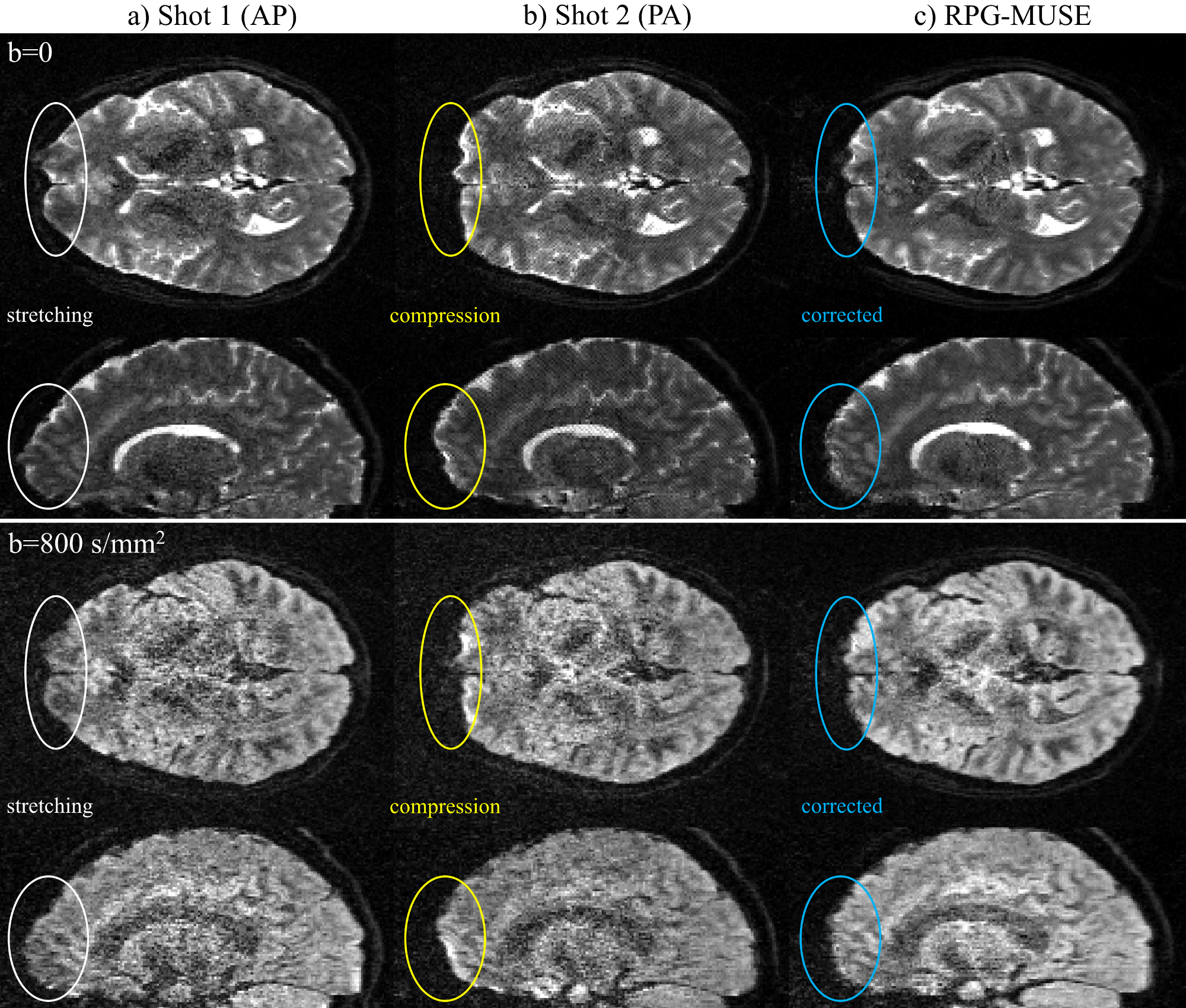
The SENSE reconstructed axial and sagittal slice images for shot 1 (PE=AP) in Fig. 2a exhibits stretching in the entire anterior region of the brain (white ovals) for both b=0 and b=800s/mm2 volumes, while the corresponding slice image for shot 2 (PE=PA) in Fig. 2b conversely exhibits compression in the same region (yellow ovals). With B-field distortions estimated from the b=0 and b=800/mm2 volumes individually in Figs. 2a and 2b, RPG-MUSE simultaneously reconstructs all shot images into a single image while correcting the distortions observed in this region (blue ovals). Derived from a combination of all four shots, the RPG-MUSE reconstructed images in Fig. 2c have an increased SNR when compared to the SENSE reconstructed shot images in Figs. 2a-b, and the improved fidelity achieved through inherent shot-to-shot motion and distortion corrections provides enhanced anatomical detail and accuracy.Conclusion
In this study, a conventional ms-EPI sequence used in acquiring dMRI data with MUSE was modified to apply alternating phase encoding gradient polarities to odd/even shots. When acquired in this manner, the RPG-MUSE algorithm presented here simultaneously accounts for shot-to-shot motion artifacts as well as geometric distortions that arise from B0 and eddy current induced B-field inhomogeneities. With such an acquisition, the overall scan time is equivalent to that of a conventional MUSE acquisition, with no need to acquire redundant data in the form of either additional b=0 volumes with RPG at the beginning of a dMRI scan1,2,3, or an entire dMRI acquisition repeated with RPG. Additionally, as the effects of eddy currents on B-field inhomogeneities are dependent on the direction of the diffusion gradients applied, this technique is able to more accurately estimate and account for the distortion characteristics associated with each diffusion volume than through distortion characteristics estimated from b=0 volumes only.Acknowledgements
This work was supported in part by NIH grants R01-NS-075017, R01- NS-074045 and R24-106048, and a grant from GE Healthcare.References
1. Bowtell R, et al. Correction of geometric distortion in echo planar images. Proc. Intl Soc. Magn. Reson. Med. 1994, p 411.
2. Andersson JLR, Skare S, Ashburner J. How to correct susceptibility distortions in spin-echo echo-planar images: application to diffusion tensor imaging. NeuroImage, 2003; 20(2):870-888.
3. Smith SM, et al. Advances in functional and structural MR image analysis and implementation as FSL. NeuroImage, 2004; 23(S1):208-219.
4. Chen NK, Guidon A, Chang HC, Song AW. A robust multi-shot scan strategy fot high-resolution diffusion weighted MRI enabled by multiplexed sensitivity encoding (MUSE). Neuroimage, 2013; 72:41-47.
5. Pruessmann KP, et al. SENSE: Sensitivity Encoding for Fast MRI. Magn. Reson. Med. 1999; 42:952-962.
6. Jenkinson M, et al. FSL. Neuroimage, 2012; 63:782-790.
Figures