5320
Non-CPMG PROPELLER diffusion imaging: comparison of phase insensitive preparation with split acquisition1Siemens Shenzhen Magnetic Resonance Ltd., Shenzhen, China
Synopsis
Turbo spin echo (TSE) based diffusion weighted (DW) PROPELLER imaging has the advantage of reduced sensitivity to B0 inhomogeneity and T2 decay caused image blurring, comparing to DW-EPI sequence. However, the violation of CPMG condition is a challenge for DW-PROPELLER sequences. Phase insensitive preparation and split acquisition methods can be used to handle the non-CPMG problem. A comparison study of these two methods was performed and the results show that the phase insensitive method has superior image quality while split acquisition method has the advantage of speed and lower SAR.
PURPOSE
Although EPI is the most common sequence for diffusion imaging due to its high data acquisition rate and insensitivity to bulk motion and physiologic movement, it suffers from image blurring caused by signal decay in the long acquisition window and static field related problems, e.g., geometric distortion and susceptibility artifact. On the other hand, the TSE based DW-PROPELLER sequence has the advantage of reduced sensitivity to B0 inhomogeneity and T2 decay caused image blurring [1]. However, CPMG conditions are hard to fulfill given that even minimums motion during application of diffusion sensitizing gradients introduces spatially varying and unknown phases to the spins in transversal plane. A phase cycling scheme was used to mitigate this problem by the DW-PROPELLER, Turboprop/Turboprop+ and X-PROP techniques. This solution requires the use of 180° refocusing pulse, so at high field SAR is high even EPI readout is applied to reduce the number of refocusing RF pulses. Besides, B1 field variation may result in unstable image quality. Another class of methods eliminate unwanted single or separate the CPMG component to avoid destructive phase interference. This kind of methods can use low refocusing angles and are not sensitive to B1 variations. This study compares two methods of the latter kind, the phase insensitive preparation method [2] and split acquisition [3].METHODS
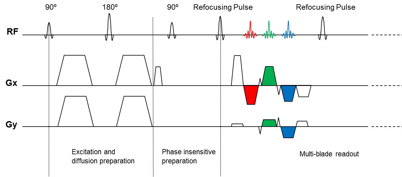
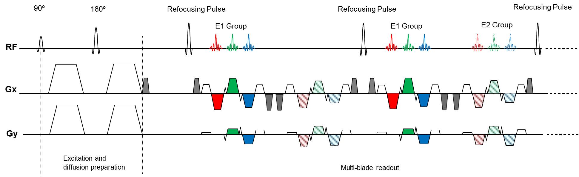
The diagrams of the proposed DW-PROPELLER sequence with phase insensitive preparation or split acquisition are shown in figure 1 and 2.
For phase insensitive preparation, a phase insensitive preparation module is inserted between the diffusion preparation and readout. In this method, a dephasing gradient is applied in the readout direction so that each voxel has equal CPMG and non-CPMG components. Then a 90° pulse with specified phase rotates the non-CPMG component to the longitudinal axis where it will be invisible in the subsequence acquisition. In addition to the dephasing gradient and 90 ° pulse, rephasing gradients must be added after each refocusing pulse and then corresponding rewinding gradients are added before the next refocusing pulse. To minimize the echo spacing, the rephasing and rewinding gradients are combined with the pre-phasing and re-phasing gradient of EPI readout, respectively.
For the split acquisition, the signal components with different phases are separated to two groups (E1 and E2). In order to mitigate the signal oscillations of the beginning echoes, a dephasing gradient right after the end of diffusion preparation is applied. Additional rephasing and dephasing gradients must be added before and after the EPI readout, respectively [4].
For both of these two sequences, multi-blade [5] acquisition strategy is applied to sampling these echoes.
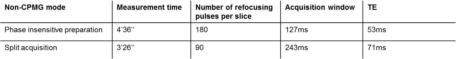
The prototype of the sequence was implemented on a Siemens MAGNETOM Aera 1.5T scanner and in vivo whole-brain data covering the whole brain were collected with both diffusion insensitive preparation and split acquisition method. The main difference of acquisition parameters of these two methods are shown in table 1.
RESULTS
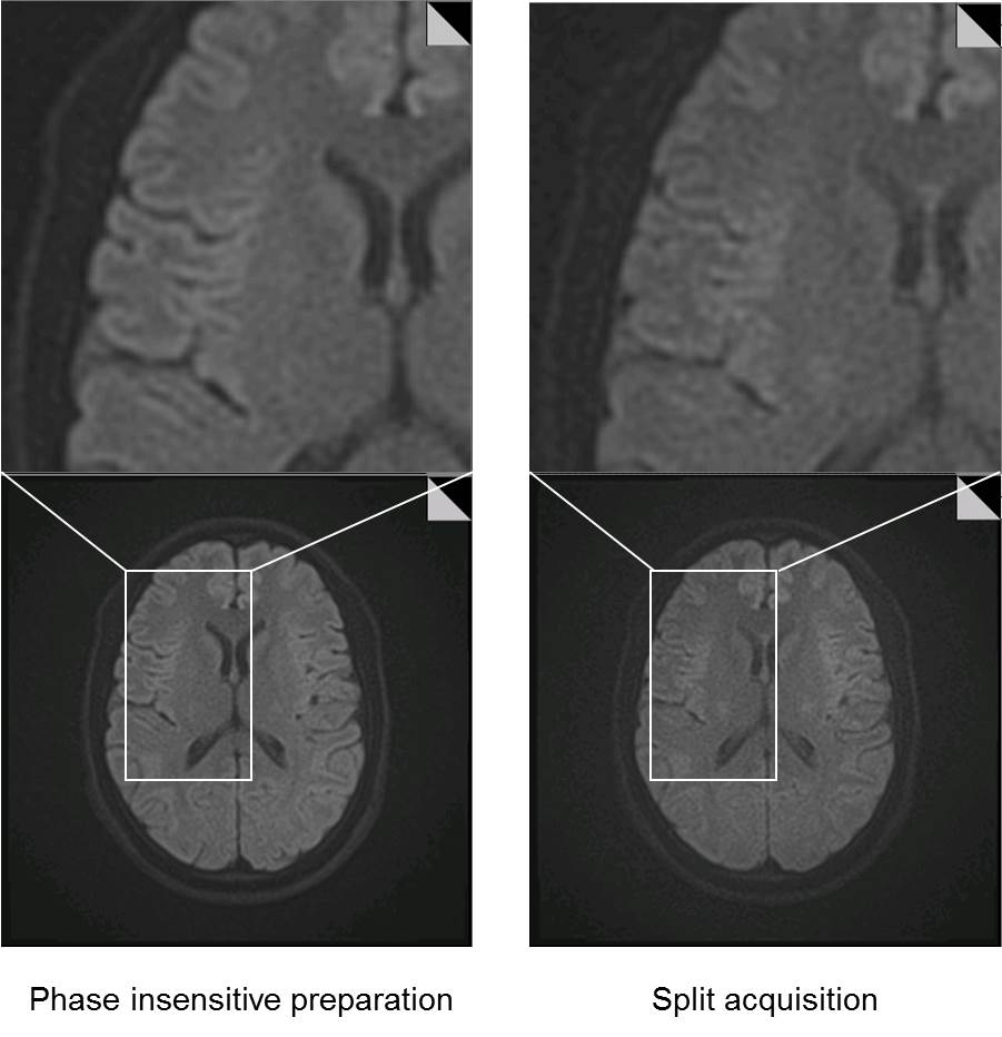
1 out of 20 slices with b = 1000 s/mm2 are shown in figure 3. The images acquired with phase insensitive preparation and split acquisition methods are shown in left and right columns, respectively. Both of these images are free of distortion and susceptibility artifacts common to DW-EPI acquisition. Besides, no intensity variations caused by violation of CPMG condition are observed on either of these two images.
The benefits of short echo train length of phase insensitive separation method can be seen as higher SNR and less image blurring. The short TE in phase insensitive method contributes to SNR as well.
The blades acquired per shot in split echo mode are twice as many as that in the phase insensitive preparation method, results in shorter acquisition time and significantly reduced RF power deposition, as shown in table 1.
DISCUSSION AND CONCLUSION
Both of the phase insensitive preparation and split acquisition methods are robust for overcoming the non-CPMG problems. Short acquisition window in phase insensitive method leads to superior image quality in terms of SNR and PSF. On the other hand, with the potential for faster acquisition with less RF power deposition, split acquisition method can be more attractive at high field.Acknowledgements
No acknowledgement found.References
1. Pipe JG. Split-blade PROPELLER DWI. In Proceedings of the 11th annual meeting of ISMRM, Toronto, Canada, 2003. p 2126.
2. Alsop DC. Phase insensitive preparation of single-shot rare: application to diffusion imaging in humans. Magn Reson Med 1997; 38: 527–533.
3. Schick F. SPLICE: Sub-Second Diffusion-Sensitive MR Imaging Using a Modified Fast Spin-Echo Acquisition Mode. Magn Reson Med 1997; 38: 638-644.
4. Zhou K, Liu W. Multi-Blade Acquisition of Split Turbo Spin Echoes: A Robust and Fast Diffusion Imaging Technique. ISMRM 24th Annual Meeting, #1821.
5. Li Z, Pipe JG, Lee CY, Debbins JP, Karis JP, Huo D. X-PROP: a fast and robust diffusion-weighted propeller technique. Magn Reson Med 2011;66:341–347.
Figures