5319
Distortion-free diffusion imaging with SMS PROPELLER DUO1GE Healthcare, Waukesha, WI, United States
Synopsis
Although fast spin echo based diffusion-weighted imaging methods provide distortion-free images, they suffer from prolonged scan times. In this work, we incorporated simultaneous multi-slice (SMS) techniques into PROPELLER DUO for accelerated distortion-free diffusion imaging. Results show that SMS PROPELLER DUO can reduce scan time dramatically which can make it a feasible option for clinical applications.
Introduction
In diffusion-weighted imaging (DWI), single-shot echo planar pulse sequences (SS-EPI) are widely used as they provide fast diffusion imaging with reduced motion artifacts. However, SS-EPI suffers from image distortions due to off-resonance effects. Fast spin echo (FSE) based sequences have been proposed to reduce image distortions in DWI [1-4]. One of the challenges in FSE-based DWI is that diffusion weighting gradients violate CPMG condition, which results in destructive interference of spin-echo and stimulated echo and unstable signal throughout the echo train. PROPELLER DUO has been proposed to mitigate these non-CPMG issues by splitting the stimulated and spin-echo coherence pathways [4]. Moreover, PROPELLER DUO has reduced scan time significantly compared to other FSE-based PROPELLER methods [4]. However, PROPELLER DUO scan time is still significantly longer than DW EPI, which limits its usage in clinical applications. Simultaneous multi-slice (SMS) techniques have been shown to reduce scan time without SNR penalty seen in conventional parallel imaging techniques [5]. To the best of our knowledge, SMS techniques have not been applied to PROPELLER DWI yet. Here, we studied the feasibility of accelerating PROPELLER DWI with SMS techniques.Methods
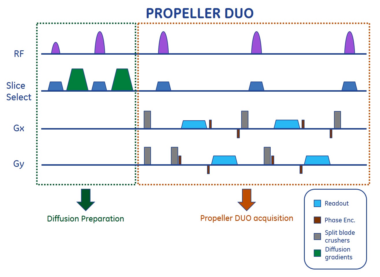
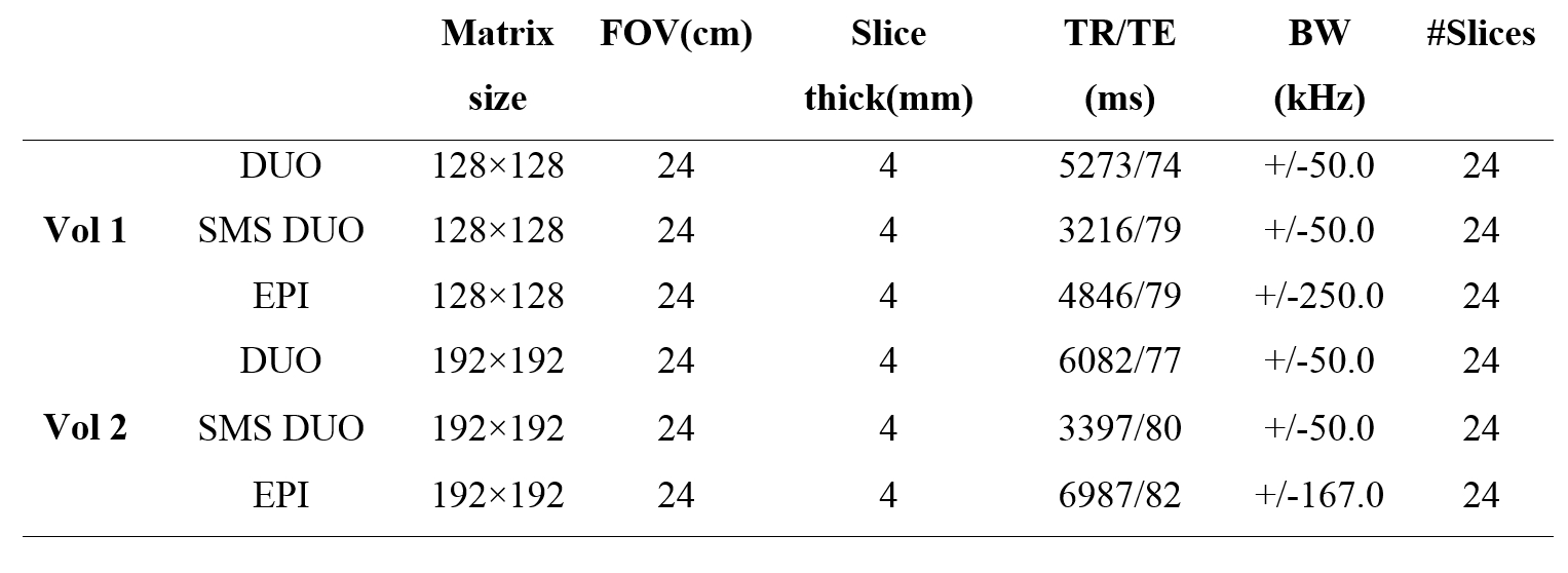
Pulse sequence diagram for PROPELLER DUO is shown in Fig. 1. SMS PROPELLER DUO is implemented using multi-band (MB) RF pulses and blipped-CAIPIRINHA [5]. Two volunteers were scanned on a 3T GE MR750W scanner using a 19-channel head coil. The study was approved by the IRB and written consents were obtained from the volunteers. Acquisition parameters are summarized in Table 1.
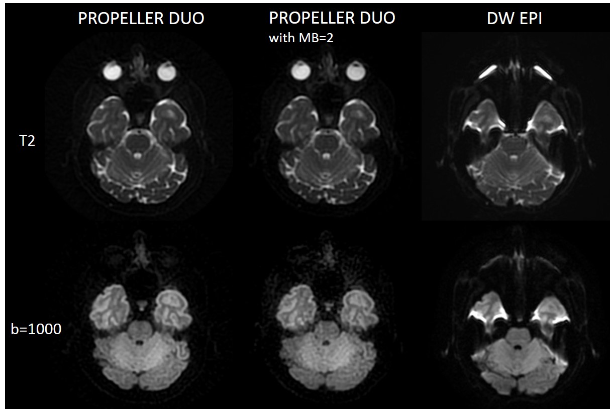
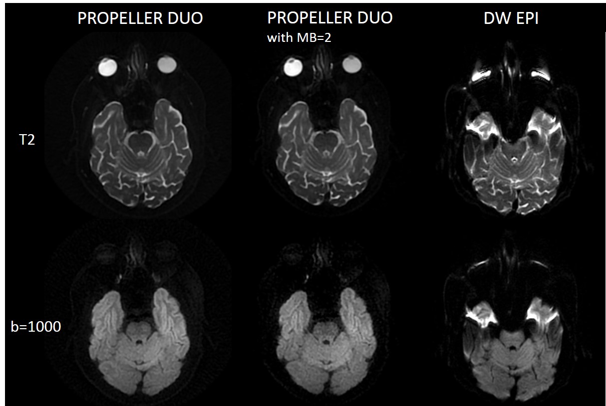
For the scans with 128×128 matrix size, total scan times were 3:46 min and 2:15 min for PROPELLER DUO and PROPELLER DUO with MB-factor of 2, respectively. For the scans with 192×192 matrix size, total scan times were 6:11 min and 3:31 min for PROPELLER DUO and PROPELLER DUO with MB-factor of 2, respectively. Slice-GRAPPA method was used for separating simultaneously acquired slices [5]. PROPELLER DUO data without MB-factor were used for calibrating GRAPPA kernels.
Results and Discussion
Fig. 2 illustrates 128×128 PROPELLER DUO images with and without MB-factor as well as DW EPI images and Fig. 3 shows 192×192 PROPELLER DUO images with and without MB-factor as well as DW EPI images. It can be seen that PROPELLER DUO images are free of image distortions observed in DW EPI images. These figures also demonstrate that SMS PROPELLER DUO with a MB-factor of 2 can reduce scan time by almost 40% without introducing artifacts in the reconstructed images. Compared to conventional RF pulses, SMS RF pulse durations get longer due to peak B1 limits and this results in increased echo-spacing. This can be seen as slight SNR reduction in SMS PROPELLER DUO images. However, using VERSE pulses can reduce peak B1 power, and therefore, reduce echo-spacing and scan time compared to non-VERSE SMS pulses [6]. In this study, PROPELLER DUO data without MB-factor were used for calibrating GRAPPA kernels, which is not feasible in clinical applications. However, it was shown that GRAPPA-kernels can also be calibrated from a separate calibration scan which can be acquired only in couple of seconds [7].Conclusion
In this study, the feasibility of SMS PROPELLER DUO is demonstrated. Preliminary results show that incorporating SMS into PROPELLER DUO can reduce scan time significantly without degrading the image quality. This reduction in scan time makes PROPELLER DUO more feasible for clinical applications.Acknowledgements
No acknowledgement found.References
1. Alsop DC. Phase insensitive preparation of single-shot RARE: application to diffusion imaging in humans. MRM 38: 527–533 (1997).
2. Pipe JG, Farthing VG, Forbes KP. Multishot diffusion-weighted FSE using PROPELLER MRI. MRM 47: 42–52 (2002).
3. Deng J, Omary RA, Larson AC. Multishot diffusion-weighted SPLICE PROPELLER MRI of the abdomen. MRM 59: 947–953 (2008).
4. Zhao X, Li Z, Gaddipati A. PROPELLER DUO: Applied to Diffusion Weighted Imaging. ISMRM 17th Annual Meeting & Exhibition (2009).
5. Setsompop K, Gagoski BA, Polimeni J, Witzel T, Wedeen VJ, Wald LL. Blipped CAIPIRINHA for simultaneous multislice echo planar imaging with reduced g-factor penalty. MRM 67: 1210-1224 (2012).
6. Conolly S, Nishimura D, Macovski A, Glover G. Variable-rate selective excitation. JMR 78:440–458 (1988).
7. Norbeck O, Mårtensson M, Avventi E, Engström M, Skare S. Self-Calibrated Simultaneous Multi-Slice PROPELLER. ISMRM 23rd Annual Meeting & Exhibition (2015).
Figures