5317
The effects of illness duration on white matter connectivity in drug-naive schizophrenia1Huaxi MR Research Center (HMRRC), Department of Radiology, West China Hospital of Sichuan University, Chengdu, China, 2Center for Information in BioMedicine, Key Laboratory for Neuroinformation of Ministry of Education, School of Life Science and Technology, University of Electronic Science and Technology of China, Chengdu, China, 3Laboratory of Cognitive Neuropsychology, Department of Medical Psychology, Anhui Medical University, Hefei, China
Synopsis
This study investigated the topological alterations of white matter connectivity in schizophrenia patients with a long illness duration by using diffusion tensor imaging and graph theoretical analysis and explored the relationship of such characteristics with the duration. We recruited three groups including the healthy controls, drug-naive schizophrenia patients with a short illness duration (0.1 to 10 months) and a long duration (12 to 36 months), and found that only the patients with a long illness duration exhibited decreased connection strength than the controls and a correlation between the nodal degree of rolandic operculum and the duration, suggesting a
Introduction
Previous studies provided evidence for abnormal brain structure in schizophrenia, while longitudinal studies further suggested that there may be a progression of these effects early in the illness (1). However, most longitudinal studies are conducted with patients treated with antipsychotic medications, which may adversely impact brain anatomy and confound explorations for disease effects over the illness course. Furthermore, the changes of the brain structural network of schizophrenia along with the illness duration have not been explored. Therefore, in order to address how different the white matter network may be altered under the long-term course of schizophrenia, the present cross-sectional study investigated the topological alterations of white matter network in never-medicated schizophrenia patients with a long illness duration.Methods
Sixty-seven drug-naive patients (aged 23.7±5.5 years) with a short illness duration (0.1 to 10 months) and 38 patients (aged 23.1±5.8 years) with a long illness duration (12 to 36 months) and 83 matched healthy controls (aged 24.6±5.1 years) were recruited. The diagnoses of schizophrenia were determined using the structured clinical interview for DSM-IV. Duration of untreated illness was evaluated by the Nottingham Onset Schedule (2), which was defined as the time period from the onset of first psychotic symptom to the date of research assessment.
All the subjects were scanned using a 3T GE MRI system. The DTI images were obtained using a single-shot EPI sequence with 15 non-collinear diffusion sensitization gradients (b=1000 s/mm2). The fiber tracking was performed in native diffusion space using the Fiber Assignment by Continuous Tracking algorithm and proceeded until either it turned an angle greater than 45° or the fractional anisotropy was less than 0.2. We used the AAL atlas to parcellate the gray matter into 90 non-cerebellar anatomical regions and obtained an FA weighted symmetrical anatomical 90 × 90 matrix for each subject.
Graph theoretical analyses were carried out using the Brain Connectivity Toolbox. A small-world network has a similar shortest path length (Lnet), but higher clustering coefficient (Cnet) than a random network (γ=Cnet/Crandom, λ=Lnet/Lrandom), and the small-worldness scalar (σ)=γ/λ will be larger than 1. The nodal degree (Si) quantifies the extent to which a node is relevant to the graph, and the total connection strength (Snet) of a network is defined as the sum of Si for all nodes in the network.
We defined a network cost threshold (from 0.1 to 0.19 with 0.01 interval). The area under the curve was calculated to provide an overall value. Among three groups, the differences in age, γ, λ, σ, Snet and Si were examined with one-way ANOVA in SPSS. Post hoc pairwise t-tests with Bonferroni correction for multiple comparisons were performed if ANOVA yielded significant results. We investigated the relationship between the topological characterizations with significant group differences and illness duration in the two patients groups respectively.
Results
All three groups showed a small-world organization of white matter networks expressed by σ>1 with γ>1 and λ≈1. These overall organization did not differ among the patients and controls (P=0.571 for σ; P=0.7 for γ; P=0.119 for λ), suggesting an intact overall organization of white matter networks in schizophrenia.
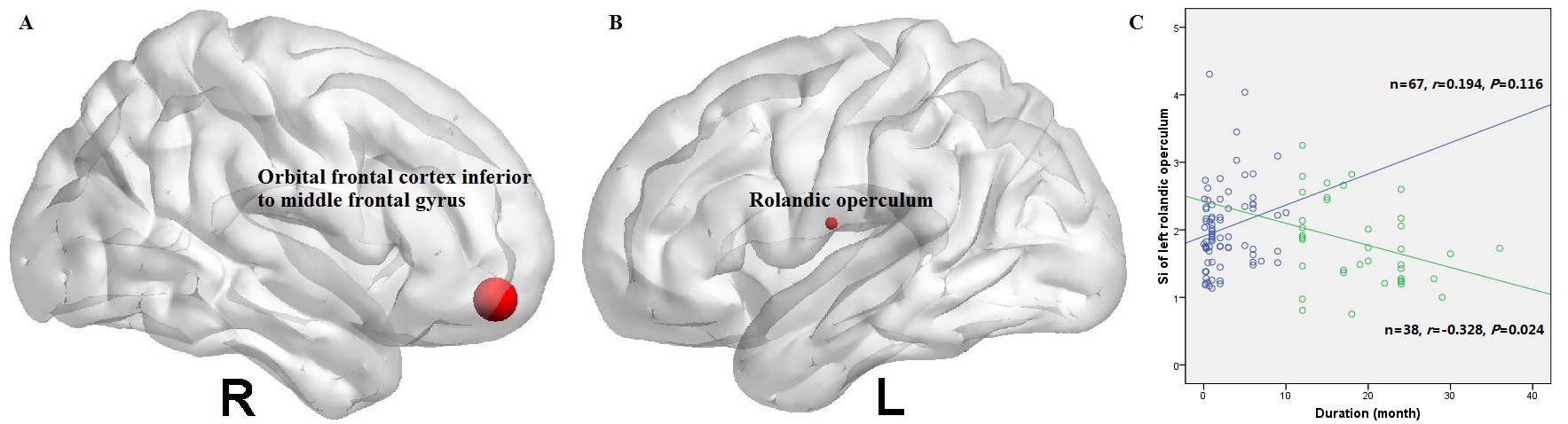
Significant group effects in the Snet was observed among the three groups (P=0.015), with decreased Snet only in patients with a long illness duration than controls (P=0.012). We further found the right orbital frontal cortex (OFC) inferior to middle frontal gyrus (P=0.001) and left rolandic operculum (P=0.008) with altered nodal degree among the three groups (Figure 1A and B). Post hoc tests revealed that the right OFC had reduced nodal degree both in patients with a short (P=0.016) and long (P=0.004) illness duration, but the left rolandic operculum exhibited reduced nodal degree only in patients with a long illness duration than controls (P=0.006). Furthermore, in the group of patients with a short illness duration, there were no significant correlations between the duration and the topological characteristics. While in the group of patients with a long illness duration, there was a significantly negative correlation between the duration and the nodal degree of the left rolandic operculum (Figure 1C).
Discussion and Conclusion
In the group of drug-naive schizophrenia patients with a long illness duration, we found a decreased structural connection strength, suggesting the shift from an optimal small-world property to a less effective organization. The left rolandic operculum negatively relating to the duration of schizophrenia with a long, but not short, illness duration suggests that this impairment may progress over the early course of illness, facilitating the understanding of the effects of illness duration on regional brain area in the white matter network of schizophrenia.Acknowledgements
No acknowledgement found.References
1. Andreasen NC, Nopoulos P, Magnotta V, et al. Progressive brain change in schizophrenia: a prospective longitudinal study of firstepisode schizophrenia. Biol Psychiatry 2011; 70:672–679.
2. Singh SP, Cooper JE, Fisher HL, et al. Determining the chronology and components of psychosis onset: the Nottingham Onset Schedule (NOS). Schizophr Res. 2005;80:117–130.
Figures