5314
Meta-analytic investigations of grey matter alterations in patients with anorexia nervosa.Simin Zhang1, Weina Wang1, Xiaorui Su1, Qiyong Gong1, and Qiang yue1
1HMRRC,Department of Radiology,West China Medical School of Sichuan University, Chengdu, China
Synopsis
The purpose of the present Meta-analytic study was to summarize the grey matter volumetric alterations and elucidate how the changes were associated with symptoms and pathophysiology in anorexia nervosa(AN). We use effect-size signed differential mapping (ES-SDM) to conduct meta-analytical whole brain volumetric differences between patients AN and healthy controls (HCs). The studies showed volume reduction in bilateral median cingulate cortex(MCC), posterior cingulate cortex (PCC), precuneus, supplementary motor area(SMA) and left cerebellum, which provide evidence for abnormalities in emotion regulation, behavior regulation and sensorimotor area in nervosa anorexia.
Objective
Anorexia nervosa is an important cause of physical and psychosocial morbidity in adolescent girls and young adult women [1]. The illness is characterized by restricted eating, an extreme fear of gaining weight and becoming fat, and often accompanied by a distorted body image. In recent years, many imaging studies have reported brain structural alterations in Anorexia nervosa, but the results are inconsistent and discrepant, therefore the neural basis of this phenomenon is not well understood yet. The current meta-analysis first summarizes voxel based morphometry findings to elucidate how the changes were associated with symptoms and pathophysiology in AN. Additionally, we investigate the influencing factors of these brain alterations which appear to be important for the prediction of the long-term outcome.Methods
A systematic search strategy was used to identify relevant whole brain voxel-based morphometry studies of patients with AD in relation to comparison groups. Relevant databases were searched for studies published between January 1994 and February 2017 using combinations of the terms “VBM”, “voxel-based morphometry” ,“whole brain” , “morphometric” and “anorexia nervosa” , “AN” or “eating disorder ”. Effect-size signed differential mapping (ES-SDM) was applied to analyze the grey matter differences between all AD patients and healthy controls. Meta-regression was used to explore the effects of demographics and clinical characteristics.Results
The studies using voxel-based morphometry image analysis were included 16 studies reported on a total of 292 AN patients and 301 healthy controls. The pooled meta-analysis did reveal significantly reduced volumes in the bilateral median cingulate cortex(MCC), posterior cingulate cortex(PCC), precuneus, supplementary motor area(SMA) , left cerebellum and right rolandic perculum in the AN patients compared with the healthy controls. No GM volume increases were found. In meta-regression analyses body mass index (BMI) in each study was negatively correlated with reduced grey matter in the bilateral precuneus.Conclusion
This meta-analysis indicates that AD patients have significantly reduced grey matter in MCC, PCC, extending to precuneus and SMA, indicating an important role of these area in the pathophysiology of the disorder. As we know, these specific regions mainly associated with emotion and sensorimotor regulation, the results fit well with the previous smaller meta-analysis by Titova et al. (2013)[2]. Additionally, volume changes in the cerebellum may also play a potentially role in AN. According to literature, the cerebellum is relevant for habit formation and ritualistic and stereotypical behavior[3], this might be associated with the often rigid and obsessive–compulsive traits in AN. Besides, the precuneus has been implicated in high-level cognitive functions, including self-related processing and aspects of consciousness[4], the meta-regression results perhaps reveal the structural underpinning of the BMI differences in pathophysiology of AD. In summary, these neural changes may underlie the clinical symptoms and pathophysiology in AN and provide the direction of future research.Acknowledgements
No acknowledgement found.References
- 1. Zipfel, S., et al., Anorexia nervosa: aetiology, assessment, and treatment. Lancet Psychiatry, 2015. 2(12): p. 1099-111.
- 2. Titova, O.E., et al., Anorexia nervosa is linked to reduced brain structure in reward and somatosensory regions: a meta-analysis of VBM studies. BMC Psychiatry, 2013. 13: p. 110.
- 3. Schmahmann, J.D., J.B. Weilburg, and J.C. Sherman, The neuropsychiatry of the cerebellum - insights from the clinic. Cerebellum, 2007. 6(3): p. 254-67.
- 4. Cavanna, A.E., The precuneus and consciousness. Cns Spectrums, 2007. 12(7): p. 545.
Figures
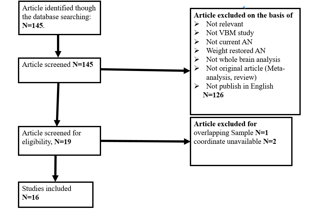
Fig. 1. Search
strategy used for the inclusion of the studies considered in the current
meta-analysis.
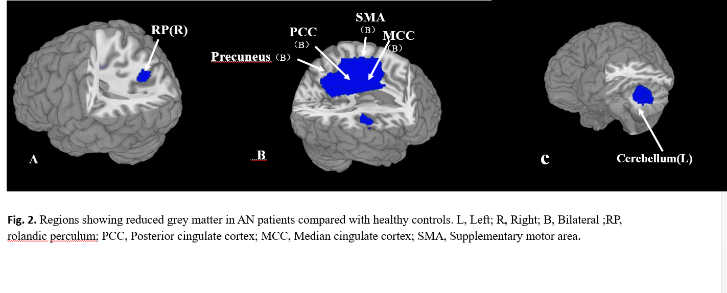
Fig. 2. Regions
showing reduced grey matter in AN
patients compared with healthy controls. L, Left; R, Right;
B, Bilateral ;RP, rolandic perculum;
PCC, Posterior cingulate cortex; MCC,
Median cingulate cortex; SMA, Supplementary motor area.
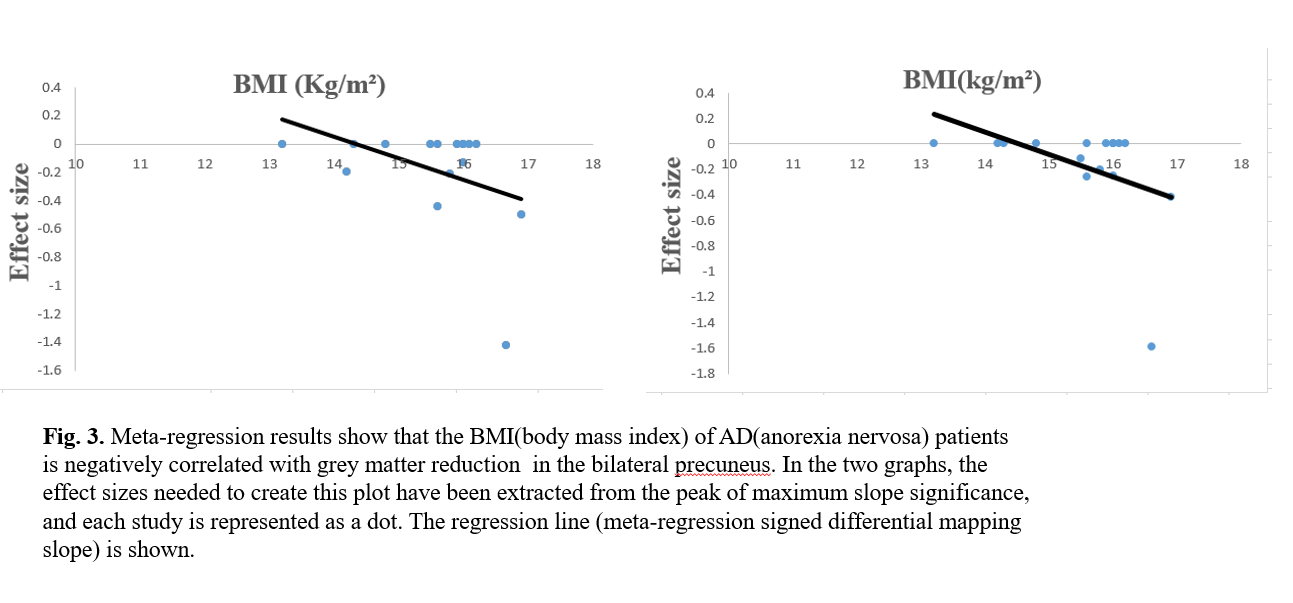
Fig. 3.
Meta-regression
results show that the BMI(body
mass index) of AD(anorexia
nervosa) patients
is negatively correlated with grey matter
reduction in
the bilateral precuneus. In
the
two graphs, the effect sizes needed to create
this plot have been extracted from
the peak of maximum slope significance, and each study is represented as a dot.
The
regression
line (meta-regression signed differential
mapping slope) is shown.
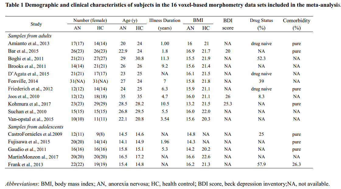
Table 1 Demographic
and clinical characteristics of subjects in the 16 voxel-based morphometry data
sets included in the meta-analysis.
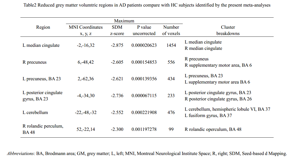
Table2 Reduced grey matter volumtric regions in AD patients compare with HC subjects identified by the present meta-analyses