5313
Altered Brain Activity in Jet Lag by Regional Homogeneity (ReHo): A resting state fMRI study1West China Hospital of Sichuan University, Chengdu, China, 2West China Hospital, Sichuan University, Chengdu, China
Synopsis
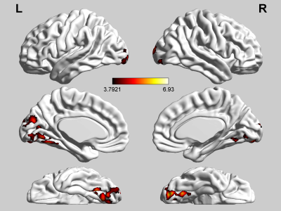
To identify how jet lag influence brain activity in rest we calculate ReHo values of 23 adult participants who were on a transmeridian flight across eight-time zones from west to east .Participants in ‘Jet Lag’ state compare to recovered state showed decreased ReHo value in the right inferior parietal lobule (BA40, BA7) and the right angular gyrus and increased in the bilateral occipital lobe. Acute circadian disruption caused by jet lag can lead to mild temporary visual cognitive dysfunction.
Background
People experience day and night alternation every and their body have the similar circadian with the environment. Jet lag happens when this balance is broken suddenly and leading to short time physical and mental symptoms. But few is known about the changes in brain caused by acute circadian disruption. We made this resting state functional MRI to identify how jet lag influence brain activity in rest。Methods
Regional homogeneity (ReHo) values of 23 adult participants who were on a transmeridian flight across eight-time zones from west to east were calculated from rs-fMRI. The data was collected the time they arriving China and two months later they recovered. All were processed and analyzed using DPARSF based on SPM8 and REST. Paired t-test was performed on individual ReHo maps between ‘Jet Lat’ state and recovered state. The relationship between ReHo value alteration and stress related hormone were analyzed.Results
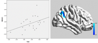
Participants in ‘Jet Lag’ state compare to recovered state showed decreased ReHo value in the right inferior parietal lobule (BA40, BA7) and the right angular gyrus. The inferior parietal lobule plays a crucial role in many higher cognitive processes, as visuo-motor transformations, maintenance and control of attention 1. The angular gyrus is one of the parts of the inferior parietal lobule and involved in visuospatial awareness 2. Decreased activation in this area may related to visual cognitive impairment. Increased ReHo value was found in the bilateral occipital lobe (including BA18, BA19, lingual gyrus and middle occipital gyrus) compare to normal state (Fig.1). The occipital lobe has the function of visual processing. Impairment of occipital lobe can lead to visual disorder accompanied with memory deficits and motor disturbance 3. Increased activity in this region may a compensatory response. The variation of ReHo in the right inferior parietal lobule has positive correlation with the variation of thyroid stimulating hormone (Fig.2) (p=0.021) and no line relationship was found in age, gender and education.Limitations
Small sample size in the current study that may affect the statistical analysis and findings. No data before the participants experience Jet Lag and further studies may can add a group not experience Jet Lag in the recent two months to identify a more rigorous result.Conclusions
Acute circadian disruption caused by jet lag can lead to mild temporary visual cognitive dysfunction. People engaged in high demand and dangerous work should have several days to recover.Acknowledgements
This study was supported by the National Natural Science Foundation (Grant Nos. 81771718, 81761128023, 81621003, 81571637). Program for Changjiang Scholars and Innovative Research Team in University (PCSIRT, Grant No. IRT16R52) of China.References
1. Mainero C CF, Pozzilli C, Pisani A, Pestalozza I, Borriello G, Bozzao L, Pantano P. fMRI evidence of brain reorganization during attention and memory tasks in multiple sclerosis. Neuroimage. 2004;21(3):858-867.
2. Ganis G, Thompson WL, Kosslyn SM. Brain areas underlying visual mental imagery and visual perception: an fMRI study. Brain research. Cognitive brain research. Jul 2004;20(2):226-241.
3. J.NewmanValérieBiousse JA. Disorders of the optic tract, radiation, and occipital lobe. Handbook of Clinical Neurology. 2011;102:205-221.
Figures

