5305
Multivariate pattern analysis of DTI reveals differential white matter in depressive patients with and without suicidal ideation.1Radiology, Huaxi MR Research Center (HMRRC), West China Hospital of Sichuan University, Chengdu, China, 2Nuclear Medicine, Huaxi MR Research Center (HMRRC), West China Hospital of Sichuan University, Chengdu, China
Synopsis
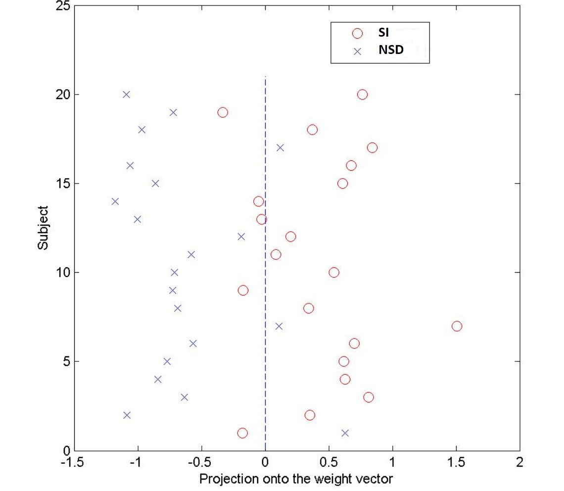
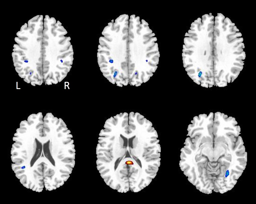
At present there are no objective biological markers that can be used to reliably identify depressive individuals with and without suicidal ideation (SI). DTI data were obtained from 20 depressive patients with SI and 20 depressive patients without SI, scanned using a 3T MRI system. Fractional anisotropy (FA) values of white matter between patients were examined using multivariate support vector machine (SVM). SVM applied to FA images correctly discriminated two groups of patients with a sensitivity of 75% and a specificity of 85% resulting in a statistically significant accuracy of 80% (p≤0.001). The discriminating regions contain the bilateral occipital lobes and parietal lobes, right temporal lobe and splenium of corpus callosum. These results reveal patterns of neuroanatomical alterations that could be used to inform the identification of depressive patients with and without SI at the individual level.
Introduction
Depression is a common psychiatric disorder affecting many people globally, and the worst outcome is suicide. At present there are no objective, biological markers that can be used to reliably identify depressive individuals with and without suicidal ideation (SI). Neuroimaging studies published so far have revealed white matter microstructure changes in patients with suicidal ideation or behavior. These studies used standard mass-univariate analytical approaches which based on average differences at the group level and therefore these results are of little use in clinical practice at the level of individual. The objective of this study was to investigate whether the application of diffusion tensor imaging could discriminate the depressive patients with and without SI at the level of individual.Methods
DTI data were obtained from 20 depressive patients with SI and 20 depressive patients without SI (non-suicidal depression, NSD), scanned using a 3T MRI system. Differences in fractional anisotropy (FA) values of white matter between patients with and without SI were examined using a multivariate pattern classification technique known as support vector machine (SVM). The accuracy of the algorithm and its statistical significance were estimated using permutation testing.Results
SVM applied to FA images correctly identified depressive patients with SI from depressive patients without SI with a sensitivity of 75% and a specificity of 85% resulting in a statistically significant accuracy of 80% (p≤0.001)(Figure 1). The discriminating white matter regions of FA contain the bilateral occipital lobes and parietal lobes, right temporal lobe and splenium of corpus callosum (Figure 2).Discussion
This study demonstrated that depressive patients with SI can be distinguished from depressive patients without SI using FA images extracted from DTI data with high classification accuracy. This classification was driven by a distributed pattern of white matter alterations which included bilateral prefrontal, occipital and parietal white matter. Multiple white matter microstructural abnormalities related in suicidality have been reported before1-3. However, previous studies used mass-univariate analyses that tend to detect only a few isolated regions with abnormal FA at group level. Here we used SVM to examine whether the whole-brain pattern of white matter microstructural abnormalities could be used to discriminate between depressive patients with and without SI at the individual level. Unlike mass-univariate analyses, SVM takes inter-regional correlations into account, and provides numerical indicators for group membership without multiple comparison biases4. Here a region's discriminative power depends not only on between-group differences in its absolute values, but also on any between-group differences in its structural correlations with other regions; this analytical approach may be particularly suited to the investigation of mental disorders such as depression or suicide behavior and ideation in which abnormalities are distributed across the whole brain. In the present study discrimination was based not only on corpus callosum but also on parts of the occipital and parietal white matter, areas not traditionally implicated in suicidal ideation; this demonstrates the ability of SVM to detect subtle and distributed white matter alterations. Consistent with previous neuroimaging studies, the microstructural alteration in corpus callosum may contribute to the disconnection between the two hemispheres in patients with SI. Fibers that travel through the splenium corpus callosum are thought to communicate somatosensory information between the two halves of the parietal lobe and visual center at the occipital lobe, and could be involved in the intelligence network and cognitive function5,6. Taken together, we found that splenium corpus callosum, occipital and parietal white matter regions had high discriminative values, providing further support for the involvement of these regions in suicidal ideation.Conclusion
These results reveal patterns of neuroanatomical alterations that could be used to inform the identification of depressive patients with and without suicidal ideation at the individual level, and provide preliminary support to the development of SVM as a clinical useful diagnostic aid.Acknowledgements
This study was supported by the National Natural Science Foundation (Grant Nos. 81621003, 81571637 and 81271532). The authors want to acknowledge the American CMB Distinguished Professorship Award to Dr Qiyong Gong.References
1. Jia Z, Huang X, Wu Q, et al. High-field magnetic resonance imaging of suicidality in patients with major depressive disorder. Am J Psychiatry. 2010;167(11):1381-1390.
2. Mahon K, Burdick KE, Wu J, Ardekani BA, Szeszko PR. Relationship between suicidality and impulsivity in bipolar I disorder: a diffusion tensor imaging study. Bipolar Disord. 2012;14(1):80-89.
3. Cyprien F, de Champfleur NM, Deverdun J, et al. Corpus callosum integrity is affected by mood disorders and also by the suicide attempt history: A diffusion tensor imaging study. J Affect Disord. 2016;206:115-124.
4. Orru G, Pettersson-Yeo W, Marquand AF, et al. Using Support Vector Machine to identify imaging biomarkers of neurological and psychiatric disease: A criticalreview. Neurosci Biobehav Rev. 2012; 36(4):1140–1152.
5. Hofer, S. and J. Frahm, Topography of the human corpus callosum revisited--comprehensive fiber tractography using diffusion tensor magnetic resonance imaging. Neuroimage. 2006; 32(3): 989-994.
6. Luders E, Narr KL, Bilder RM, et al. Positive correlations between corpus allosum thickness and intelligence. Neuroimage. 2007; 37(4):1457-1464.
Figures