5298
Analysis of resting-state networks alterations of first-episode obsessive–compulsive disorder by ICA -based fMRI1Magnetic resonance imaging Department, The First Affiliated Hospital of Zhengzhou University, Zhengzhou City, China, 2GE Healthcare, MR Research China, Zhengzhou, China, 3The First Affiliated Hospital of Zhengzhou University, Zhengzhou, China
Synopsis
Whole brain resting-state functional connectivity was analyzed using independent component analysis in 47 subjects including 23 first-episode and treatment-naïve patients with obsessive-compulsive disorder(OCD) and 24 health controls. Three abnormal resting-state networks, pDMN, RFP and lVN, have been found in OCD patients. In addition, OCD patients showed increased functional connection in Bilateral cuneus(T=3.8222,P=0.005), Right inferior parietal lobule(T=5.291,P=0.005)and Right middle occipital lobule(T=4.614,P=0.005) compared with controls. It’s considered that changes of abnormal resting-state networks might be associated with emotional and cognitive dysfunction in OCD patients.
Purpose
Obsessive-compulsive disorder (OCD) is a psychological disorder with an estimated lifetime prevalence of 1% to 3% 1, characterized by uncontrollable obsessive thoughts, compulsive behavior and compulsions to counteract these thoughts and behavior clinically 2. As known that, OCD is among the leading causes of functional disability worldwide, but the neurobiology and aetiological origins of it is not clear. Recently, a specific network of cortical-striatal-thalamic-cortical (CSTC) circuits has been suggested in the pathophysiology of OCD 3. However, many of the previous resting-state functional magnetic resonance imaging (rs-fMRI) studies in OCD depended on prior knowledge. Is there any other OCD-related functional changes occur besides the CSTC circuits remains unanswered. This study intend to investigate the whole brain rs-fMRI connectivity alternation of first-episode and treatment-naïve OCD patients by independent component analysis (ICA) to explore the pathophysiology of OCD .Method
A total of 47 subjects were recruited including 23 drug-naive OCD patients and 24 healthy controls with matched age, gender, handedness and education level in this study (Table1). OCD patients were diagnosed through Structured Clinical Interview for DSM-IV Axis I Disorders-Non-patient Edition (SCID-I) by an experienced psychiatric chief physician. MRI datasets were acquired on a 3T GE Discovery 750 System (GE Healthcare, Milwaukee, MI, USA) scanner equipped with an 8-channel parallel head coil. Functional MRI data were obtained via a GRE-EPI sequence (TR/TE=2000/30msec, FOV= 220×220mm ; matrix= 64× 64; flip angle= 90°; slice thickness=4.0mm, slice gap=0.5mm, 32 axial slices, flip angle=90°, 180 time points, 5760 images in acquisition , 6 minutes total time). Subjects were instructed to stay alert and relaxed with eyes closed and no body motion during imaging sessions. Standard image data preparation and pre-processing were performed with MRIcroN software (http://www.mricro.com) and DPARSFA software based on MATLAB 2012b (http://www.mathworks.com). Group independent component analysis (ICA) was using rs-fMRI image with GIFT software (http://icatb.sourceforge.net). Statistical analysis was carried out with SPM8 software (http://www.fil.ion.ucl.ac.uk/spm). One-sample T-test(FWE correction, α=0.05, p<0.05) was performed to make masks of networks component, and two-sample T -test(Alphasim correction, α=0.01, p<0.05) was performed to seek the significant difference of functional networks between OCD patients and controls.Results
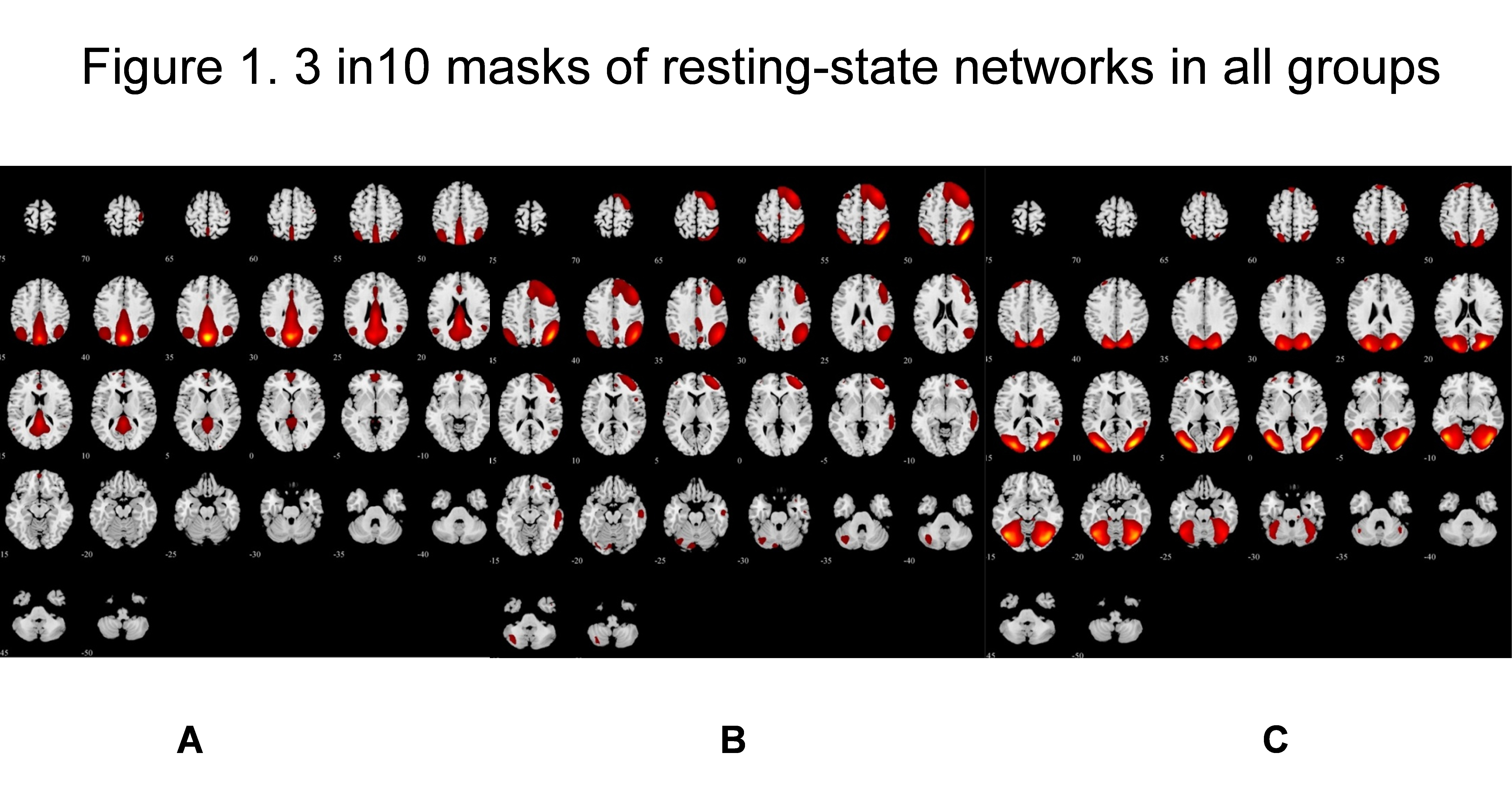
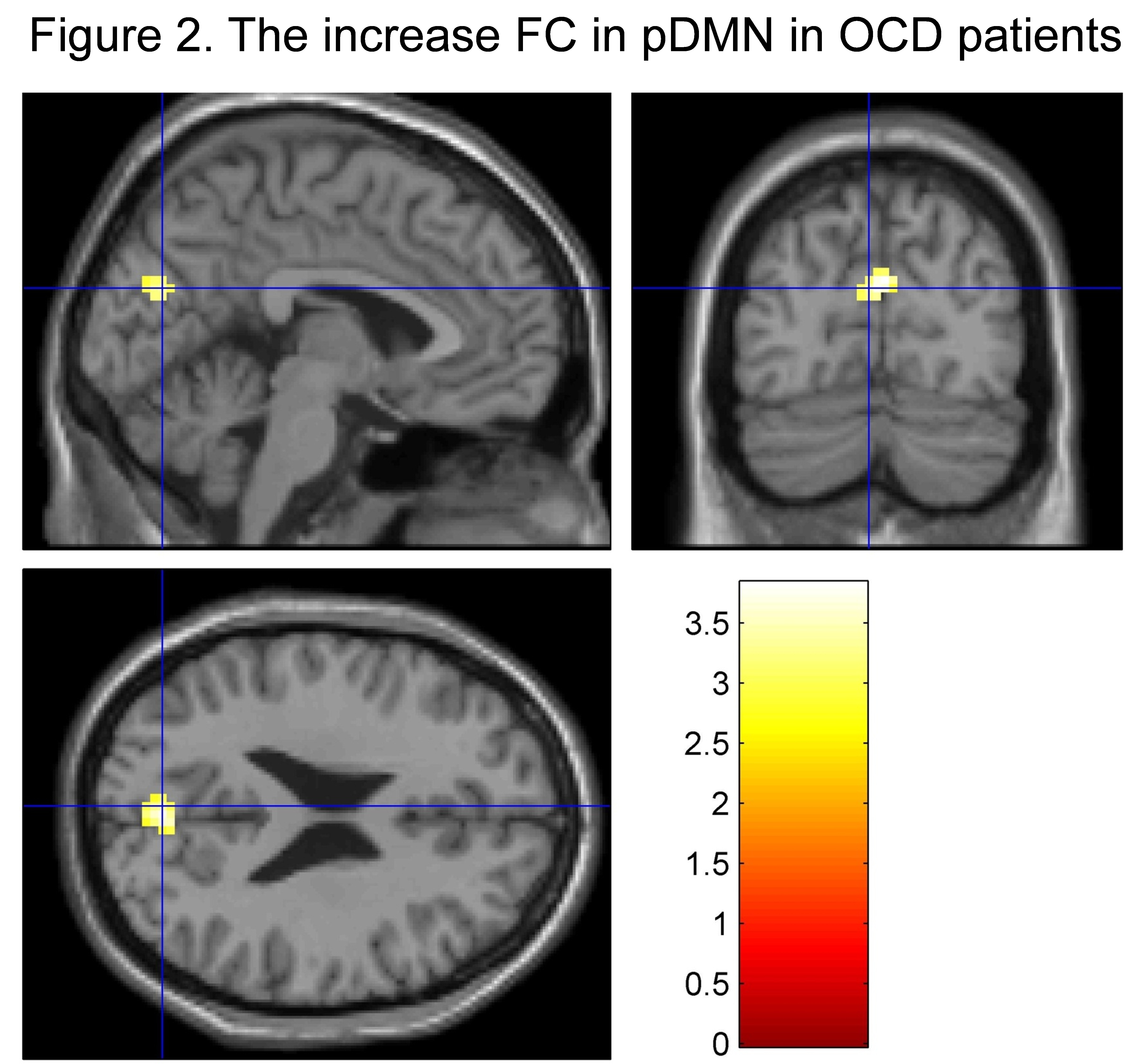
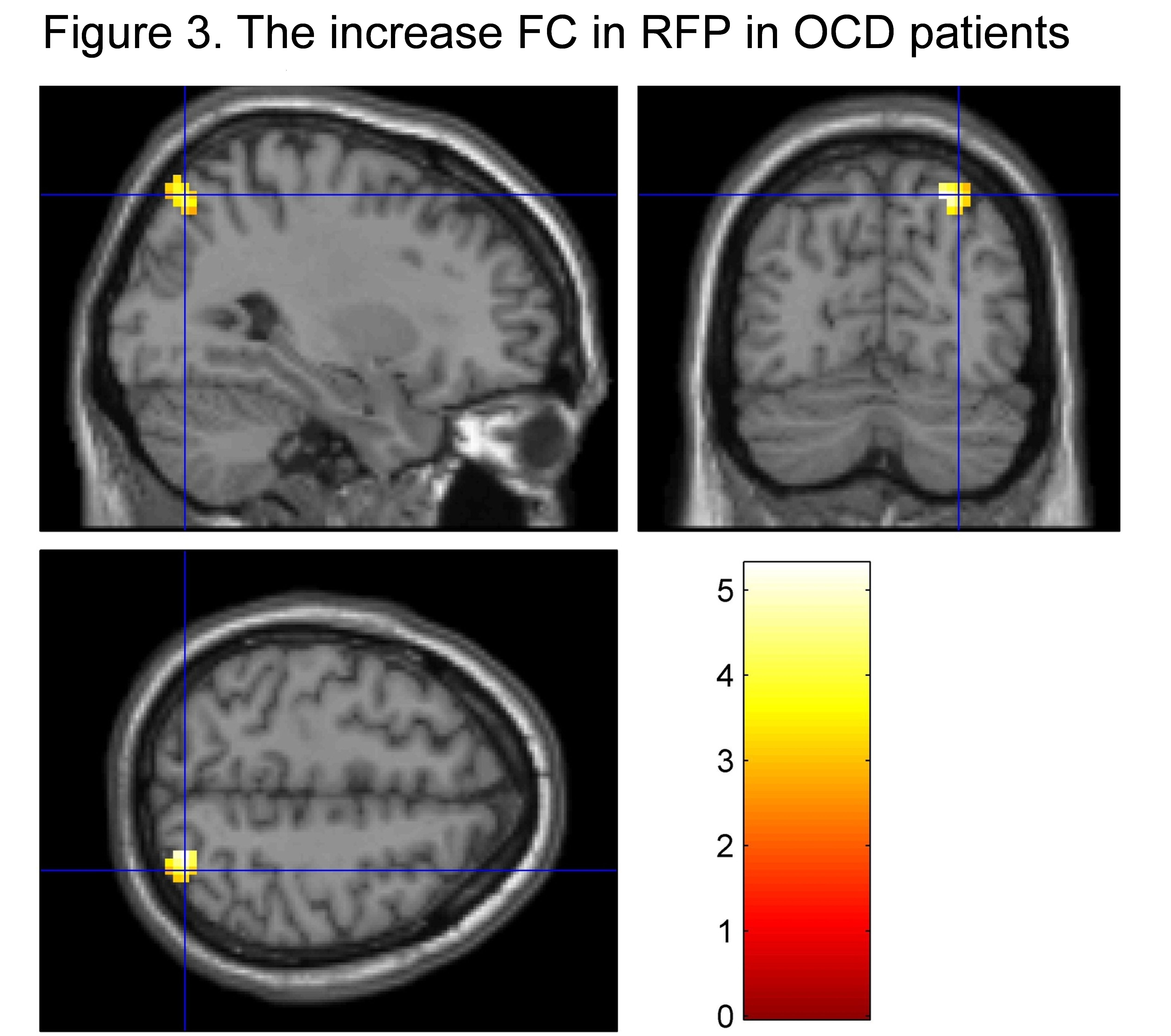
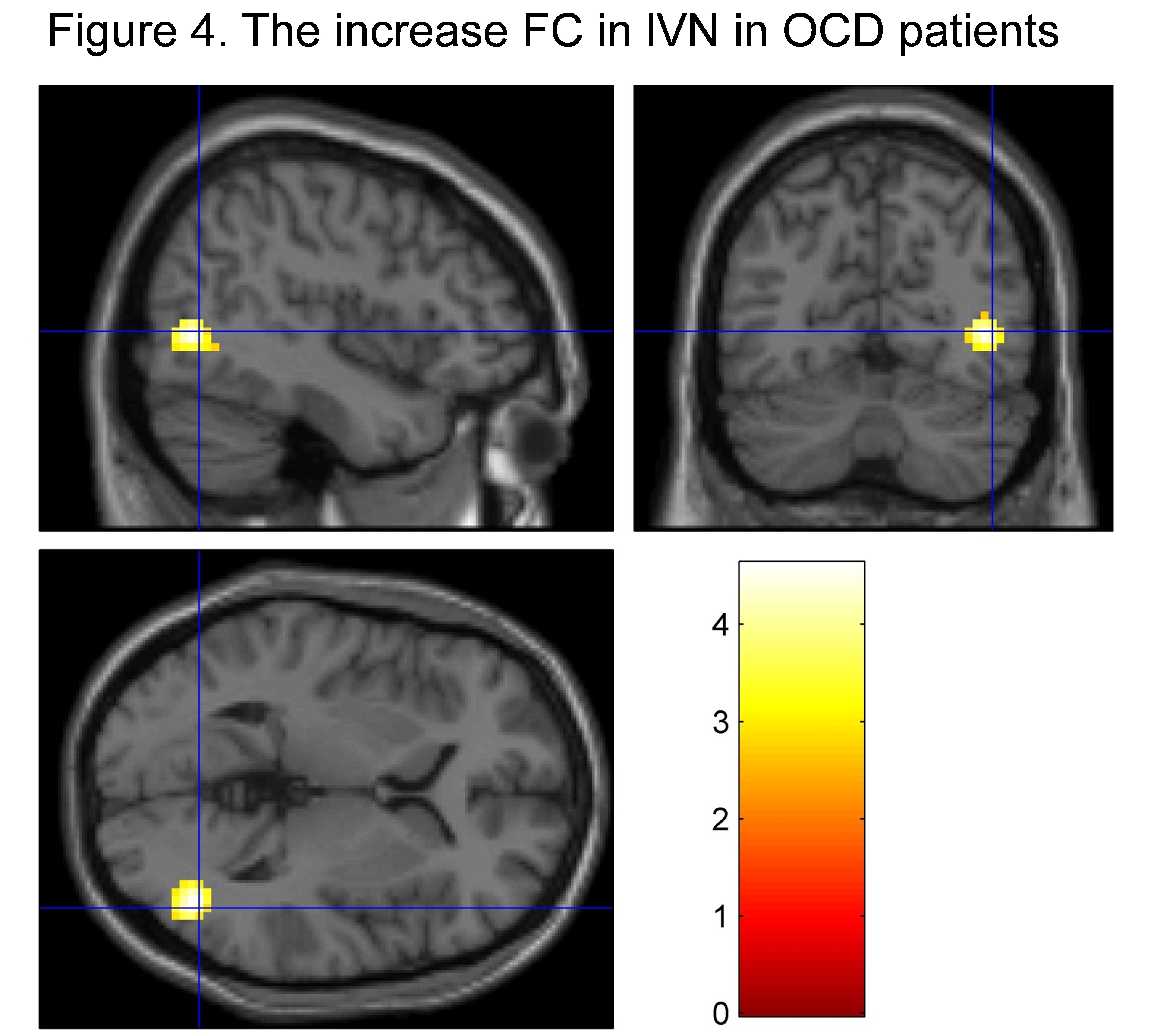
10 masks of resting-state networks were found in both groups. The resting-state networks, posterior default-mode network (pDMN), right frontoparietal network (RFP) and lateral visual network(lVN), appeared significantly difference compared with control group, were shown in Fig 1. The current results showed that the functional connectivity (FC) of pDMN network in OCD patients was higher than that in healthy controls in bilateral cuneus(T=3.822,P=0.005)(Fig 2). The FC of RFP network in the right inferior parietal lobule was higher than that in healthy controls(T=5.291,P=0.005)(Fig 3). Besides, the FC of lVN in the right middle occipital lobule was higher than that in healthy controls(T=4.614,P=0.005)(Fig 4).Discussion and Conclusion
The results of whole brain resting states functional networks in OCD patients without drug and psychosocial interventions showed more objective and reliable information for psychological mechanism of OCD. Noise was removed effectively, and altered brain connectivity between the first-episode and treatment-naïve OCD patients and health controls was found without any priori assumptions and model driven by using ICA. This study revealed that there were three abnormal resting state networks, pDMN, RFP and lVN, which mainly represented increased functional connectivity in OCD patients. Altered abnormal resting-state networks might be associated with emotional and cognitive dysfunction in OCD patients. It is believe that this noninvasive method might be useful for exploring the pathophysiology of OCD .Acknowledgements
No acknowledgement found.References
[1] Ruscio AM, et al. (2010). The epidemiology of obsessive—compulsive disorder in the National Comorbidity Survey Replicatio. Mol Psychiatry. 15:53-63.
[2] Burdick KE, et al. (2008). Neurocognitive profile analysis in obsessive compulsive disorder. J Int Neuropsychol Soc. 14:640–645.
[3] Menzies L, et al. (2008). Integrating evidence from neuroimaging and neuropsychological studies of obsessive-compulsive disorder: the orbitofronto-striatal model revisited. Neurosci Biobehav Rev. 32:525-549.
Figures