5287
DTI Analysis in FLAIR-positive Lesions and Normal-Appearing White Matter in Young Adult Multiple Sclerosis Patients1Radiology, Stony Brook Medicine, Stony Brook, NY, United States, 2Biomedical Engineering, Stony Brook University, Stony Brook, NY, United States, 3New York University, New York, NY, United States
Synopsis
Neuroimaging studies showed that there is abundant diffusion tensor imaging (DTI) research that has been done on older multiple sclerosis (MS) patients. By contrast, similar research is relatively sparse in young/pediatric MS patients. Our study is interested in discovering the effects of MS on young adult patients to see whether the same pattern as in older patients appears. Our results suggest that DTI diffusivity data provides insights in the pathophysiology of MS in young adults. Diffusivity data may serve as an imaging biomarker of early disease pathophysiology in MS.
Purpose
Multiple Sclerosis, one of the most common white matter diseases, breaks down the connections between peripheral muscular nerves and the central nervous system (CNS) through demyelination.1-2 Despite the prevalence of the disease, the cure and causes of MS still remain unknown. Many scientific studies have showed that DTI is able to suggest parameters of demyelination and axonal loss, in addition to showing a relatively good sensitivity to white matter damages in MS.2 Therefore, its noteworthy results have played a contributory role in the understanding of this disease.1-4 However, most of MS research has focused on patients over the age of 40, and similar studies on young adult MS patients are sparse. In order to gain a better understanding of how MS progresses and whether or not it has the same effects on young adult patients, this study aims to investigate axial (AD), radial diffusivity (RD) and fractional anisotropy (FA) in FLAIR-positive lesions and normal-appearing white matter in young MS patients.Methods
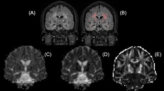
This study included 22 relapsing-remitting MS patients between the ages of 18 and 35 (25.77 ± 5.03 years) with disease duration of 0.02 to 11 years, and 17 age-, gender-matched healthy controls (22.19 ± 2.79 years). A 3T Siemens scanner was used to acquire T2 FLAIR and DTI images for each participant, and all images manipulations tools that are used in this study are ITK-Snap which was used for lesion segmentation (http://www.itksnap.org/pmwiki/pmwiki.php); FSL, which was used for images co-registration and DTI calculations, (https://fsl.fmrib.ox.ac.uk/fsl/fslwiki/FSL) and Freesurfer to perform images reconstruction(http://freesurfer.net). FLAIR positive lesions were segmented and also verified by our neuroradiologist. DTI parameters (AD, RD, FA) were tabulated and calculated for the region of interests (ROIs) of FLAIR-positive lesions in MS and normal-appearing white matter (Figure 1). Comparisons were made with corresponding regions in controls. The same process was also done in grey matte. Lesion maps were created for each patient, and were overlaid on control subjects to run a voxel based analysis in order to calculate FA differences between groups. As a result, FA differences between patients and controls were significantly enhanced in the brain. Correlations were made with EDSS scores, disease duration, lesion volume and counts as well. The statistical comparison used unpaired t-test and correlation analysis.Results and Discussions
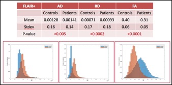
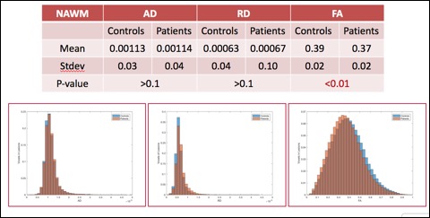
In FLAIR-positive lesions (Figure 2), AD and RD values in young/pediatric MS patients were significantly higher (P<0.005 and P<0.0002 respectively) compared to age-matched healthy controls. In NAWM lesions, RD and AD were not significantly different whereas FA was significantly different between patients and controls. In NAWM (Figure 3), although FA was significant between groups, there were significant overlaps. The differences are likely due to partial voluming with lesions.
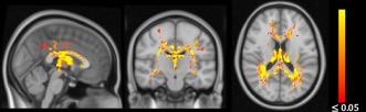
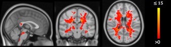
Figure 4 shows that FA was significantly different on white matter tracts within periventricular areas of the brain; and there were maximally 15 lesions located at the same area of the brain as shown in Figure 5.
In gray matter, there was no significance shown in DTI analysis between patients and controls. This could be simply that MS is a common white matter disease and early stage of the disease.
Lastly, no correlation was shown between DTI parameters and EDSS, and between DTI parameters and disease duration. We observed markedly decreased FA in MS patients compared with other similar studies done in older 5, suggesting FA changes are more prominent in young adult and pediatric-onset MS patients
Conclusion
The overall preliminary results are consistent with other published studies on older adults. Despite the consistency shown between DTI values, DTI diffusivity data can still provide insights into the pathophysiology of MS in young adults.1,2,4 Diffusivity data may serve as an imaging biomarker of early disease pathophysiology in MS. It is suggested that future studies should be longitudinal to determine any significant changes in lesions over time; and correlation between DTI and Neuropsychological Scores should be done in the future since it is beneficial to know how MS affects patients’ behaviors and cognitive disabilities.Acknowledgements
No acknowledgement found.References
1. Hasan, Khader M., et al. “Diffusion tensor fractional anisotropy of the normal-Appearing seven segments of the corpus callosum in healthy adults and relapsing-Remitting multiple sclerosis patients.” Journal of Magnetic Resonance Imaging, vol. 21, no. 6, 2005, pp. 735–743., doi:10.1002/jmri.20296.
2. Sbardella, Emilia, et al. “DTI Measurements in Multiple Sclerosis: Evaluation of Brain Damage and Clinical Implications.” Multiple Sclerosis International, vol. 2013, 2013, pp. 1–11., doi:10.1155/2013/671730.
3. Zavodszky, Maria I., et al. “Feasibility of Imaging Myelin Lesions in Multiple Sclerosis.” International Journal of Biomedical Imaging, vol. 2011, 2011, pp. 1–12., doi:10.1155/2011/953806.
4. Roosendaal, S, et al. “Regional DTI differences in multiple sclerosis patients.” NeuroImage, vol. 44, no. 4, 2009, pp. 1397–1403., doi:10.1016/j.neuroimage.2008.10.026.
5. Fink, Frauke, et al. “Comparison of Diffusion Tensor-Based Tractography and Quantified Brain Atrophy for Analyzing Demyelination and Axonal Loss in MS.” Journal of Neuroimaging, vol. 20, no. 4, May 2009, pp. 334–344., doi:10.1111/j.1552-6569.2009.00377.x.
Figures