5267
Evaluating the effect of 3-n-butylphthalide on expression of aquaporin-4 by ultra-high b value diffusion weighted imaging in animal model with focal cerebral ischemia1Department of MRI, The First Affiliated Hospital of Zhengzhou University, Zhengzhou, China, 2GE Healthcare, China, Beijing, China
Synopsis
Previous studies have reported that 3-n-butylphthalide (NBP) had beneficial effects on stroke through multiple aspects, including decreasing the area of cerebral infarct, improving energy metabolism , inhibiting the inflammatory response and improving cerebral microvessels . Although the positive effects of NBP on cerebral ischemia and cerebral infarct have been verified in ischemic patients and animal models, the effects of NBP in aquaporin-4 (AQP-4) are still unclear. Recently, ultra-high diffusion weighted imaging was reported to be able to reflect AQP-4 changes . This study would like to evaluate the effect of 3-n-butylphthalide on expression of aquaporin-4 by ultra-high b value diffusion weighted imaging in animal model of focal cerebral ischemia at different time points.
INTRODUCTION
The water channels aquaporin-4 (AQP-4) normally present in the brain are implicated in regulation of water exchanges across the cell membrane in normal and in pathological conditions 1. The l-isomer of 3-n-butylphthalide (NBP) was extracted as a pure component from seeds of Apium graveolens Linn. Previous studies have reported that NBP had beneficial effects on stroke through multiple aspects, including decreasing the area of cerebral infarct 2, improving energy metabolism 3, inhibiting the inflammatory response 4 and improving cerebral micro vessels 5. Although the positive effects of NBP on cerebral ischemia and cerebral infarct have been verified in ischemic patients and animal models, the effects of NBP in AQP-4 are still unclear. Recently, ultra-high diffusion weighted imaging was reported to be able to reflect AQP-4 6 . This study would like to evaluate the effect of 3-n-butylphthalide on expression of aquaporin-4 by ultra-high b value diffusion weighted imaging in animal model of focal cerebral ischemia at different time points. METHODS
The experimental protocol was approved by the hospital's ethics committee for animal studies under the surveillance of the National Animal Research Authority. A total of
55 male Sprague–Dawley rats weighting 260 to 305g were used in this studies. Rats were randomly divided into three groups with administered 10 ml/kg saline (MCAO
group, n=25; sham group, n=5) or 10 mg/kg 3-n-Butylphthalide (NBP group, n=25) per day by gavage for five days. The MCAO and NBP groups were subjected to transient
focal cerebral ischemia by intraluminal middle cerebral artery (MCA) blockage with a 5–0 monofilament nylon suture, as described 7. And MR scans were operated at 1,
3, 6, 12, 24h after modeling. MR Images were acquired on a 3.0T MRI scanner (Discovery MR 750; GE Medical Systems, Milwaukee, WI, USA) with a 4-channel animal coil
using a multi-b single shot SE-EPI pulse sequence with the following parameters: TR/TE=3200/101 ms, acceleration factor 2, slice thickness 3 mm with no gap and FOV
70×35, matrix size 96×64. For each subject, six b-values set at (number of excitation, NEX): 2000(5), 2500(6), 3000(8), 3500(8), and 4000(10) and 4500(10) s/mm2 in three
orthogonal directions were acquired. The ADCuh was calculated by the mono-exponential model. Five rats of the two groups at each time point and the sham group were
executed. The immunohistochemistry of AQP-4 expression was performed and integrated optical density (IOD) was measured by HPIAS-000. SPSS 17.0 software was used
to analyze the varying trends in ADCuh values and the IOD of AQP-4. The comparison among the three groups at different time points was based on one way ANOVA, with
P < 0.05 indicating statistical significance. METHODS
RESULTS
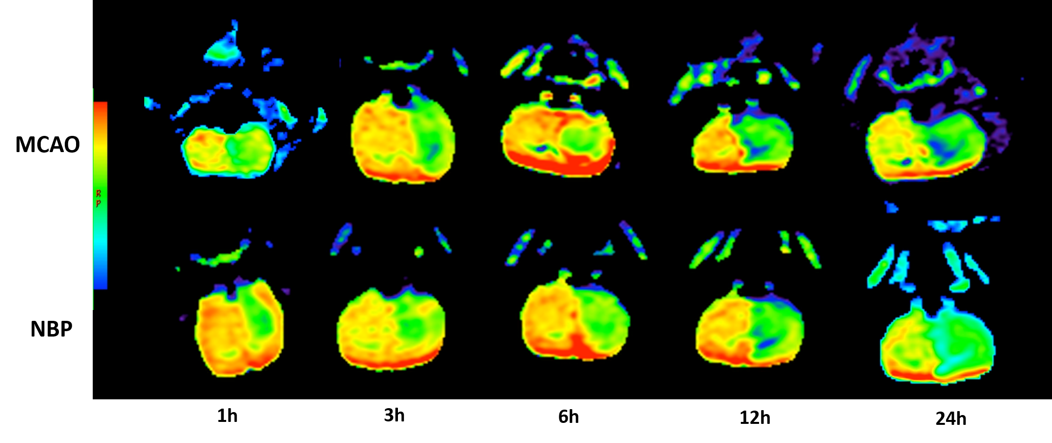
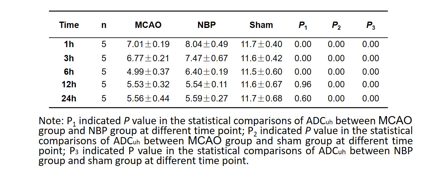
Representative ADCuh maps (Figure 1) show reduced ADCuh on the left hemisphere in the two groups after 1h onset. Significantly less reduction in ADCuh was seen in
NBP rats from 1h onset to 6h. The ADCuh in MCAO fell to the lowest at 6h, but at 12h fell to the lowest in butylphthalide. Compared to the sham group, the ADCuh in both
MCAO and NBP group appeared significantly decrease. There was no significant differences of the ADCuh value of involved side in MCAO and NBP group after 12h and
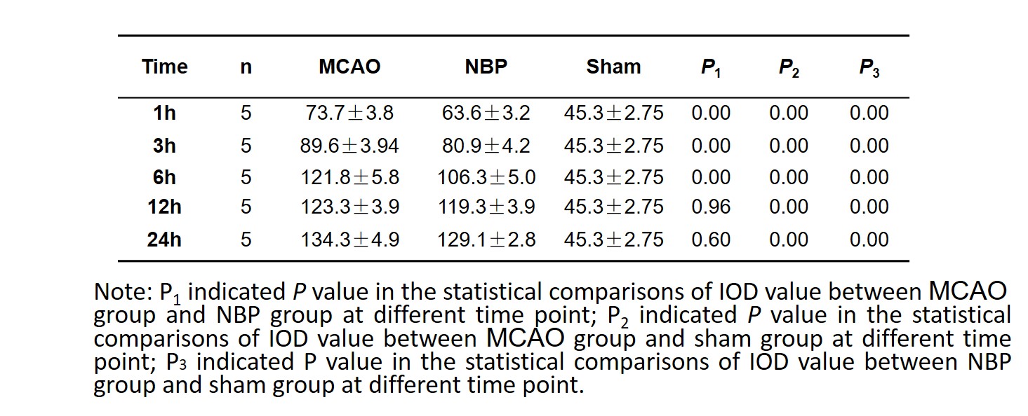
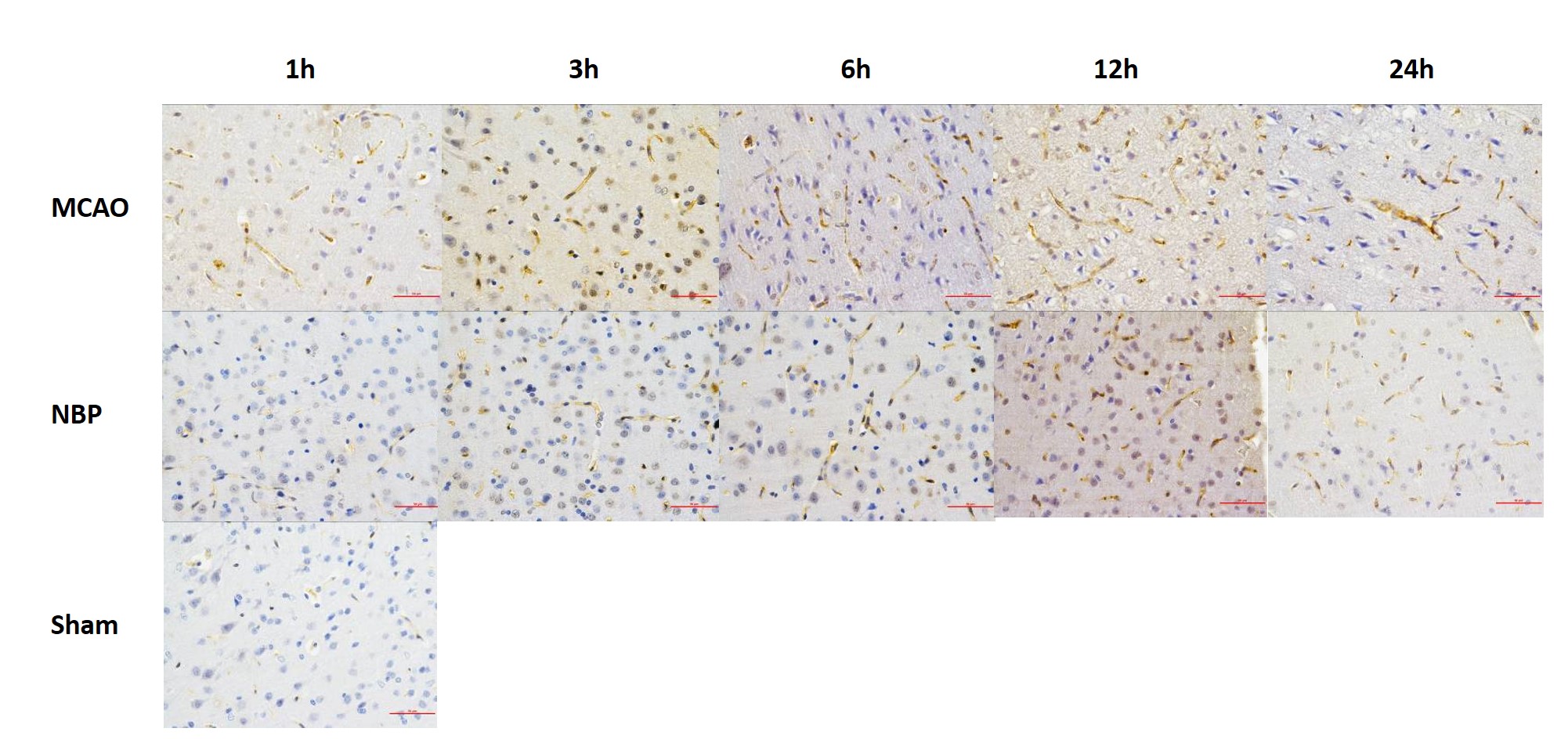
24h onset (Table 1). Histochemical micrographs of rat brain at different time points after intraluminal middle cerebral artery blockage showing AQP-4 immunolabeling
was. AQP-4 expression was seen in MCAO group and butylphthalide group after 1h, and reached the peak after 24h. And the difference between MCAO and sham、NBP
and sham was statistically significant (P2、P3<0.05). In 1-6h the difference between MCAO and NBP group was statistically significant (P1<0.05).(Table 2) RESULTS
DISCUSSION
The principal finding of this study was significantly reduced ADCuh values and AQP4-increase in both MCAO rats and rats with NBP treated. But, compared to the MCAO
group, the NBP treated rats showed less reduction, which indicated the protection effect of NBP in ischemia. ADCuh values generated by ultra-high-b-values could
eliminates the influence of microvascular perfusion and the signal intensity changes were mainly resulted from the slow diffusion component. A higher b-value DWI could
provide better contrast with its reflection of more tissue diffusivity and less T2 shine-through effect 8. The molecular cascade initiated by cerebral ischemia includes the
loss of membrane ionic pumps and cell swelling. DISCUSSION
CONCLUSION
Our study showed that the ADCuh value decreases sharply in the early stages of MCAO, and there was a negative correlation between AQP-4 expression and ADCuh values. In conclusion, ADCuh may provide some information of aquaporin molecular and butylphthalide may reduce ischemic cerebral edema through regulating AQP-4 expression.Acknowledgements
No acknowledgement found.References
1. Pallab Bhattachary, Anand Kumar Pandey, Sudip Paul, Ranjana Patnaik. Melatonin renders neuroprotection by protein kinase C mediated aquaporin-4 inhibition in animal model of focal cerebral ischemia. Life Sciences. 2014, 100, 97–109
2. Liu XG, Feng YP. Protective effect of dl-3-n-butylphthalide on ischemic neurological damage and abnormal behavior in rats subjected to focal ischemia. Yao Xue Xue Bao 1995;30:896– 903. Chinese.
3. Feng YP, Hu D, Zhang LY. Effect of DL-butylphthalide (NBP) on mouse brain energy metabolism in complete brain ischemia induced by decapitation. Yao Xue Xue Bao 1995;30:741–44. Chinese.
4. Xu HL, Feng YP. Inhibitory effects of chiral 3-n-butylphthalide on inflammation following focal ischemic brain injury in rats. Acta Pharmacol Sin 2000;21:433–8.
5. Liu CL, Liao SJ, Zeng JS, Lin JW, Li CX, Xie LC, et al. dl-3nbutylphthalide prevents stroke via improvement of cerebral microvessels in RHRSP. J Neurol Sci 2007;260:106–13.
6. A Mukherjee, D Wu, HC Davis , et al. Non-invasive imaging using reporter genes altering cellular water permeability.Nature Communications, 2016 , 7 :13891
7. G. Yang, P.H. Chan, J. Chen, E. Carlson, S.F. Chen, P. Weinstein, C.J. Epstein, H. Kamii. Human copper–zinc superoxide dismutase transgenic mice are highly resistant to reperfusion injury after focal cerebral ischemia. Stroke, 1994; 25, 165–170
8. Burdette JH, Durden DD, Elster AD, et al. High b-value diffusion-weighted MRI of normal brain. J Comput Assist Tomogr. 2001;25(4):515-19.
Figures