5251
White Matter Fiber Structure Revealed by Synthetic MRI of the Longitudinal Magnetization Relaxation Rate (R1): Effects of Age at 1.5T1Boston University, Boston, MA, United States
Synopsis
Purpose: To investigate the potential of R1-weighted Synthetic-MRI for unraveling the microstructure of white matter and for constructing accurate high resolution brain connectomes. Methods: Eighteen research subjects ranging in age from 0.6 to 87years were scanned with multispectral qMRI (T1, R1, T2, and PD) and analyzed with R1-weighted Synthetic MRI. Results: connectome renderings as function of increasing age show the expected increased WM track bundle packing and anatomical distributions evolution as function of age. Conclusion: R1-weighted WM Fibrography is a promising complementary alternative to DTI-WM Tractography for studying the microarchitecture of white matter.
Purpose
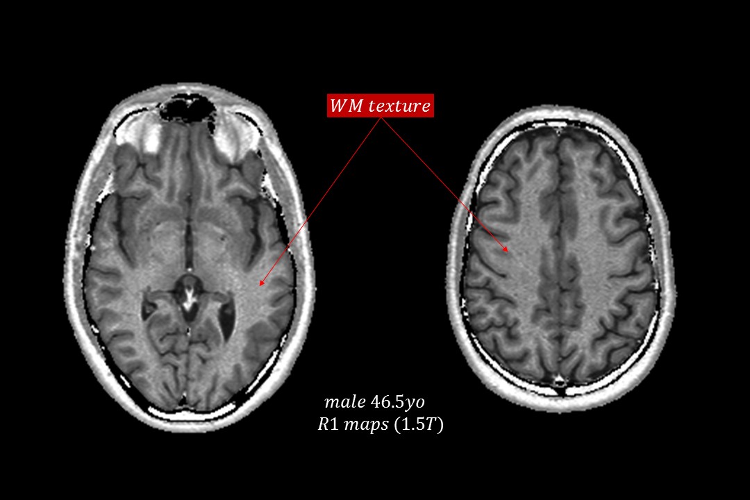
White matter (WM) typically appears as a smooth continuous tissue in MRI. One exception are qMRI maps of the longitudinal relaxation rate R1 in which WM exhibits an irregular and well-defined, but subtle graininess (Figure 1). The purpose of this work was to investigate the potential of Synthetic-MRI to further accentuate the visual appearance of this WM texture as observed on the R1 maps by means of exponential R1-weighted image synthesis. Further objectives were to assess visually the texture changes as function of age, and by using the multislice R1-weighted image data sets, to generate three-dimensional renderings of the full WM circuitry (connectome) using standard 3D-to-2D projection techniques.Methods
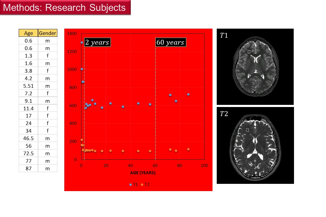
HIPAA compliant prospective study approved by the local IRB included eighteen research subjects ranging in age from 0.6 to 87years (Figure 2). Head MRIs were acquired with the mixed turbo spin echo pulse (mixed-TSE) sequence at 1.5T (Intera and Achieva, Philips Healthcare, Best, the Netherlands) using the body and head coils for RF transmit and signal receive respectively: voxel = 0.9 x 0.9 x 3 mm3. Images were qMRI processed to generate maps of T1 (and R1) and T2, and the normalized proton density (PD) using model conforming qMRI algorithms programmed in Mathcad (PTC, Needham, MA). These multispectral qMRI maps were used as virtual patients to synthesize R1-weighted images by means of the equation:
$$Synth Img(Rs)=PD exp(-Rs/ R1) $$
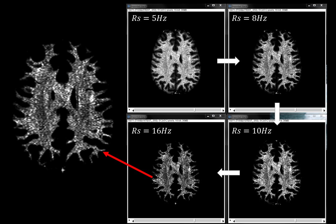
The synthesizing rate parameter Rs was varied in the range 0 to 16Hz. R1-weighted images were read with ImageJ (https://imagej.nih.gov/ij/), sharpened using the "unsharp mask" method and the connectome renderings were generated with the Volume Viewer plugging.
Results
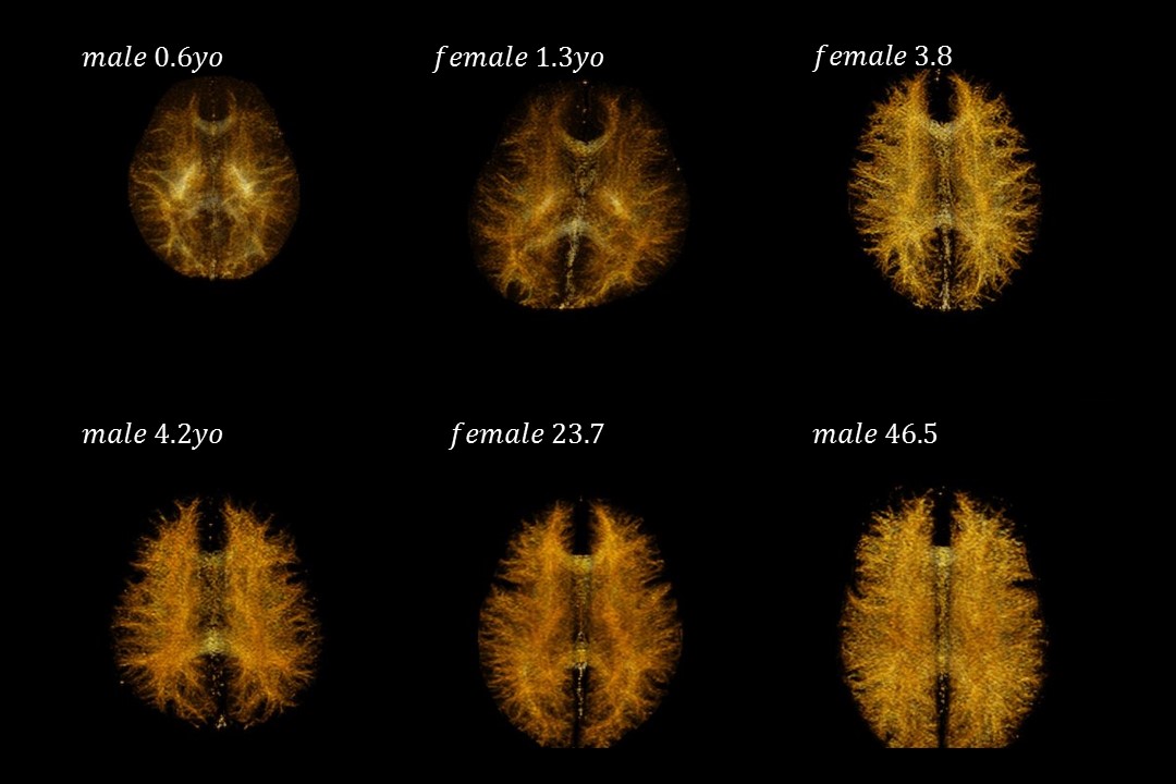
WM texture was observed in the synthetic images of the 18 subjects studied. Well-defined texture delineation was reached (Figure 3) in the range Rs>10Hz. The visually perceived “texture density” increased as a function of increasing age and these changes were most pronounced from 0.6 years to adulthood. Selected examples of connectome renderings as function of increasing age show the expected increased WM track bundle packing and anatomical distributions evolution as function of age (Figure 4).Discussion
Rendering the full brain WM circuitry with conventional diffusion tensor (DTI) MRI techniques, which use the Stejskal-Tanner diffusion encoding pulses, can pose significant technical and mathematical challenges (1,2). Furthermore, DTI infers indirectly the location and the shape of individual fiber tracts through a series of mathematical procedures and geometrical assumptions. The accuracy of the generated fiber tracts has been recently called into question by several groups (3,4). The R1-image synthetic technique described in this abstract is direct, anatomically self-evident, fast, and the gradient power can be fully utilized for spatial encoding; therefore the connectome renderings resolution and accuracy are limited solely by the native resolution of the directly acquired images and the accuracy of the PD and R1 qMRI algorithms.Conclusion
A new technique for visualizing WM and its evolution as function of age has been developed and tested at 1.5T: R1-weighted WM Fibrography via Synthetic MRI is a promising complementary alternative to DTI-WM Tractography for studying the microarchitecture of white matter
WMF can generate undistorted high spatial resolution connectomes in clinically feasible (<10min) scan times using standard clinical MRI hardware. This work could have implications for the assessment of WM disease and for improving preoperative surgical planning, and for building ultrahigh spatial resolution connectomes for use in routine clinical practice.
Acknowledgements
No acknowledgement found.References
1. McNab JA, Edlow BL, Witzel T, et al. The Human Connectome project and beyond: Initial applications of 300 mT/m gradients. NeuroImage 2013;80:234-245.
2. Fan Q, Witzel T, Nummenmaa A, et al. MGH–USC Human Connectome Project datasets with ultra-high b-value diffusion MRI. NeuroImage 2016;124:1108-1114.
3. Shawna Farquharson, J.-Donald Tournier, Fernando Calamante, et al. White matter fiber tractography: why we need to move beyond DTI. Journal of neurosurgery 2013;118(6):1367-1377.
4. Maier-Hein K, Neher P, Houde J-C, et al. Tractography-based connectomes are dominated by false-positive connections. bioRxiv 2016.
Figures