5247
White Matter Microstructural Change Following Traumatic Brain Injury Assessed by Simultaneous Multi-Slice Multi-Shell Diffusion MRI - A Preliminary Study1National Intrepid Center of Excellence, Walter Reed National Military Medical Center, Bethesda, MD, United States
Synopsis
Mild traumatic brain injury (mTBI) is difficult to diagnose and characterize. In this study, we applied simultaneous multi-slice multi-shell diffusion MRI to assess white matter microstructural changes in chronic military mTBI. Preliminary results showed parameters derived from Mean Apparent Propagator MRI method are superior to the parameters derived from diffusion tensor imaging or diffusion kurtosis imaging in differentiating tissues with distinct structural and architectural features, and thus has increased ability to identify microstructural changes in mTBI.
Introduction
Mild traumatic brain injury (mTBI) is difficult to diagnose and characterize. Identifying underlying aberrant white mater (WM) structural changes associated with persistent post-concussive symptoms can differentiate mTBI from purely psychological disorders. Advanced diffusion MRI techniques using q-space diffusion MRI show promising results in assessing brain tissue microstructure. For example, rotation invariant and scalar parameters computed from the Mean apparent propagator (MAP) MRI (Özarslan et al., 2013) show consistent variation across neuroanatomical brain regions and increased ability to differentiate tissues with distinct structural and architectural features compared with diffusion tensor imaging (DTI)-derived parameters (Avram et al., 2016). However, long scan times have limited their clinical utility. We evaluated feasibility of using simultaneous multi-slice (SMS) multi-shell diffusion MRI in assessing chronic military mTBI in this study.Method
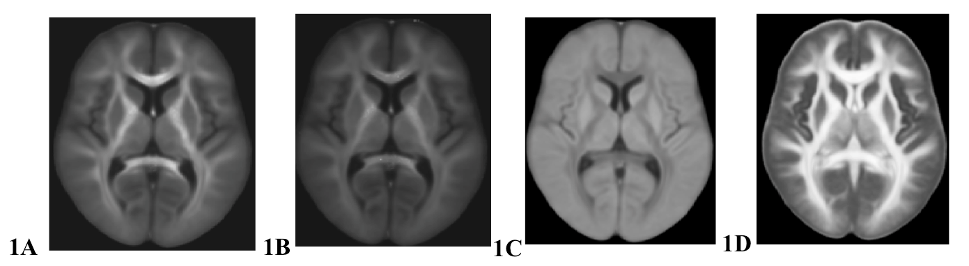
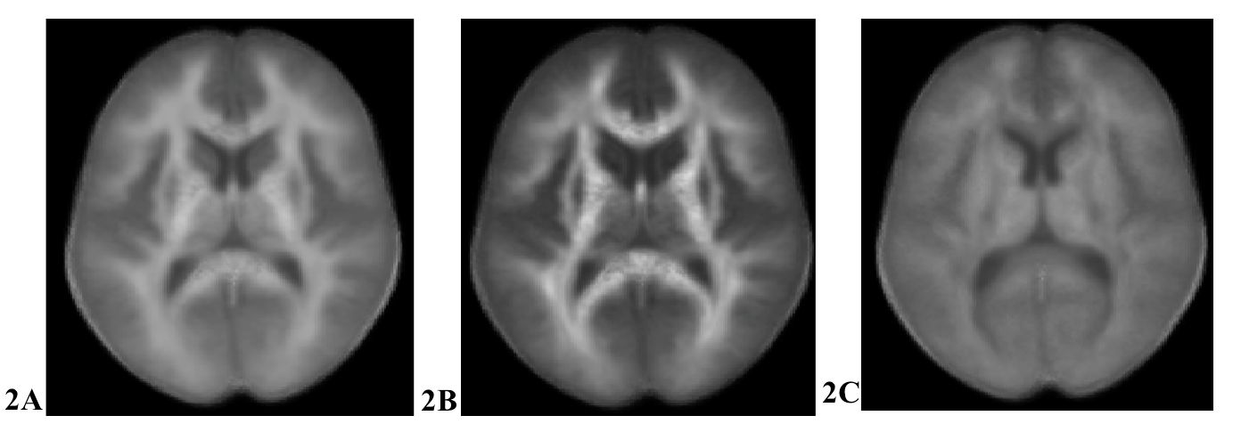
Sixty-one service members (age: 39.8 ± 4.9 years old, M/F = 59/2) previously diagnosed with mTBI received a series of neuroimaging exams at the National Intrepid Center of Excellence (NICoE) using a 3T scanner equipped with a 32-channel head coil. Nineteen non-TBI controls were recruited for comparison (age: 32.7 ± 6.7 years old, M/F = 14/5). SMS multi-shell diffusion MRI was acquired in about 25 minutes using sparse and optimal acquisition (SOA) schemes (Koay et al., 2012) with three shells (b=1000, 2000, 3000, 1.7 mm3) and a SMS acceleration factor of three. After noise reduction, motion eddy current correction and non-linear registration to the structural T2W image using the TORTOISE package (Pierpaoli, et al., 2010), MAP MRI parameters, including the return-to-origin probability (RTOP, Fig. 1A), the return-to-axis probability (RTAP, Fig. 1B), the return-to-plane probability (RTPP, Fig. 1C), and the propagator anisotropy (PA, Fig. 1D), were derived by estimating the probability density function (PDF) of spin displacements in complex microstructure (Özarslan et al., 2013). In addition the DTI-derived parameters, fractional anisotropy (FA) and mean diffusivity (MD); and the diffusion kurtosis imaging (DKI)-derived parameters, mean kurtosis (MK), axial kurtosis (AK) and radial kurtosis (RK) (Fig. 2) were estimated. General linear mixed modeling was applied to evaluate the difference of MAP MRI-derived parameters between non-TBI and mTBI subjects by modeling covariates of age and gender. Significance was tested using non-parametric permutation test with TFCE (Threshold-Free Cluster Enhancement, Smith et al. 2016) as well as Monte Carlo simulations with 0.05 family-wise error for correcting multiple comparisons of the whole brain voxel-wise analysis.Results
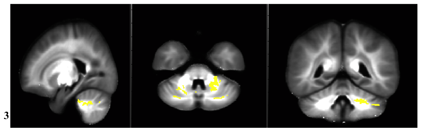
There is no significant statistical difference of DTI or DKI measures between TBI and non-TBI group after correcting for multiple comparisons, nor was there for RTOP or RTAP MAP MRI measures. Compared to non-TBI controls, the mTBI group had lower RTPP mainly over the bilateral cerebellar hemispheric white matter (Fig. 3), suggesting disrupted restricted barriers along the parallel orientation, probably caused by axonal injury.Discussion and Conclusion
In this cohort with a relative small sample size, we have demonstrated the feasibility of applying advanced diffusion MRI techniques to identify WM microstructural changes related to mTBI. The RTOP has been suggested as an indicator for restricted diffusion (Özarslan et al., 2013); while the RTAP and RTPP reflect the presence of restrictive barriers in the radial and axial orientation, respectively (Avram et al., 2016). Our findings of lower RTPP over the cerebellum and brainstem region suggest decreased restricted barriers in the axial orientation, likely caused by axonal injury in mTBI. In addition, our results showed MAP MRI is more sensitive than DTI measures in assessing WM microstructural changes in mTBI. Since our non-TBI group was younger than the TBI group, we are aware that age effects may contribute significance to these findings. For this ongoing project we will evaluate possible confounding effects by using a larger sample size. These results suggest that SMS multi-shell diffusion MRI might be sensitive to white matter changes in chronic military mTBI. In conclusion, SMS q-space imaging may have application in monitoring persistent post-concussive symptoms and brain recovery in chronic military mTBI patients.Acknowledgements
This study was supported through the Congressionally Directed MedicalResearch Program, grant DM130132.
Disclaimer: The views expressed in this abstract are those of the authors and do not reflect the official policy of the Department of Army/Navy/Air Force, Department of Defense, or U.S. Government.
References
1. Koay C.C. et al. (2012) Medical Physics. 39(5), 2499-2511.
2. Avram A.V. et al. (2016) Neuroimage 15(27):422-434.
3. Özarslan E. et al., (2013) Neuroimage 18:16-32.
4. Pierpaoli, C. et al. (2010) ISMRM 18th Annual Meeting (https://science.nichd.nih.gov/confluence/display/nihpd/TORTOISE).
Figures