5199
Ultrashort Echo Time-MRI of proximal femoral cortical bone in patients with osteoporosis1Radiology, NYU Langone Health, New York, NY, United States, 2Department of Rheumatology, NYU Langone Health, New York, NY, United States, 3Osteoporosis Center, Hospital for Joint Diseases, NYU Langone Health, New York, NY, United States
Synopsis
Ultra-short echo time MRI (uTE-MRI) allows exploration of the role of cortical bone properties in osteoporosis. OP can be primary or can also be induced by drugs such as glucocorticoids (glucocorticoid-induced osteoporosis, GIO).No previous studies have assessed cortical porosity in GIO patients compared to OP patients.Our aim was to perform uTE-MRI of cortical bone of the proximal femur (a common site of fracture) in GIO patients and compare the results to those of OP patients using a bi-exponential fitting procedure of uTE images.
Introduction:
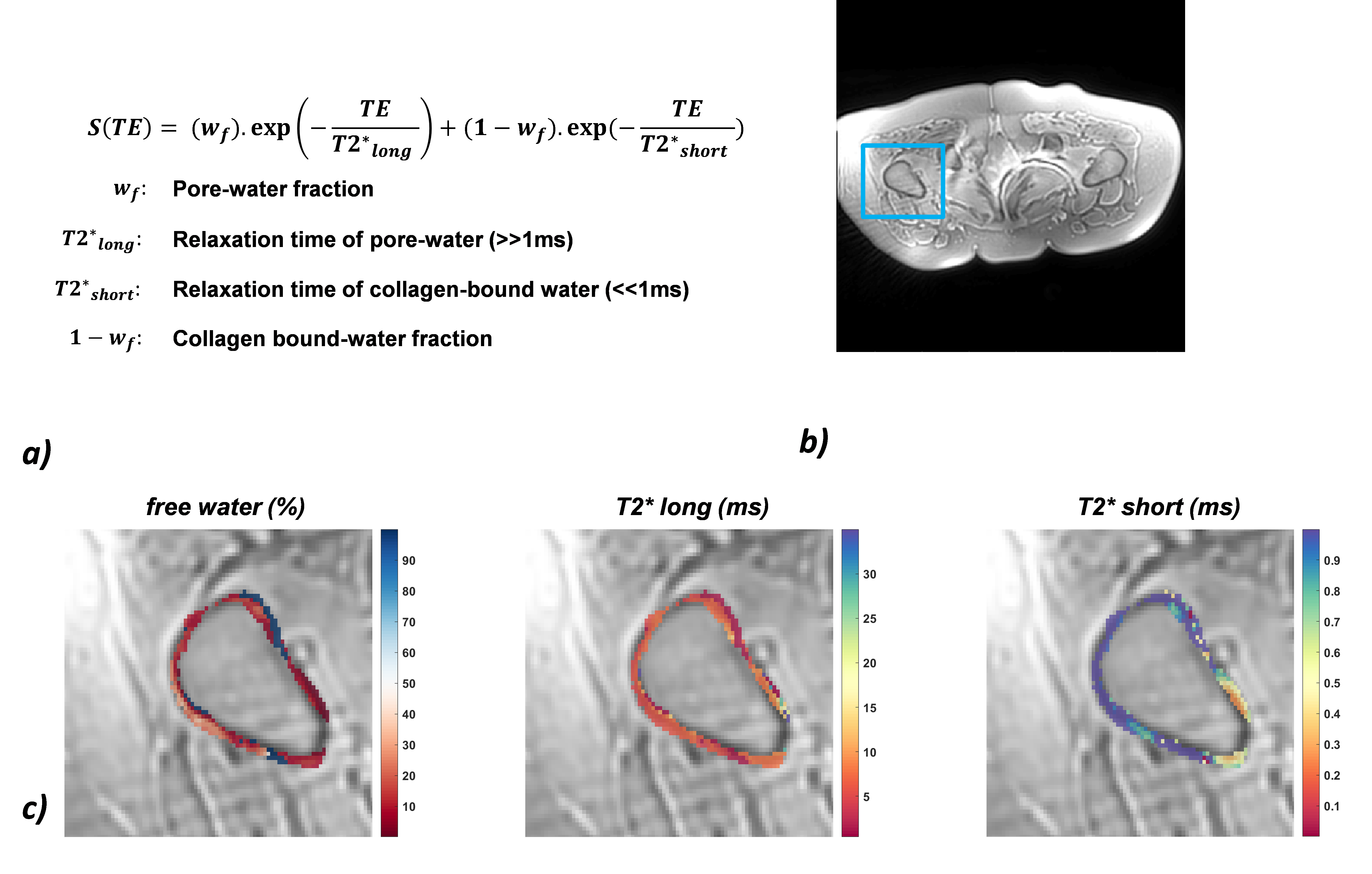
Recent developments in ultra-short echo time MRI (uTE-MRI) allows exploration of the role of cortical bone properties in osteoporosis. Previous methods have used measurement of hydrated proton collagen through bi-exponential fitting1,2 or the use of an inversion-recovery preparation module to suppress the long-T2 component3. For the study of osteoporosis (OP), such techniques can be of interest since collagen is linked to bone porosity at the cortical level4. The bi-exponential model has been studied mainly in the knee, tibia, and in-vitro showing two regimes: one ascribed to pore (free) water and its relaxation characterized by T2* long and one ascribed to collagen-bound water associated with short T2*. OP can be primary or can also be induced by drugs such as glucocorticoids (glucocorticoid-induced osteoporosis, GIO)5. No previous studies have assessed cortical porosity in GIO patients compared to OP patients. Therefore, the goal of our study was to perform uTE-MRI of cortical bone of the proximal femur (a common site of fracture) in GIO patients and compare the results to those of OP patients.Material & Method
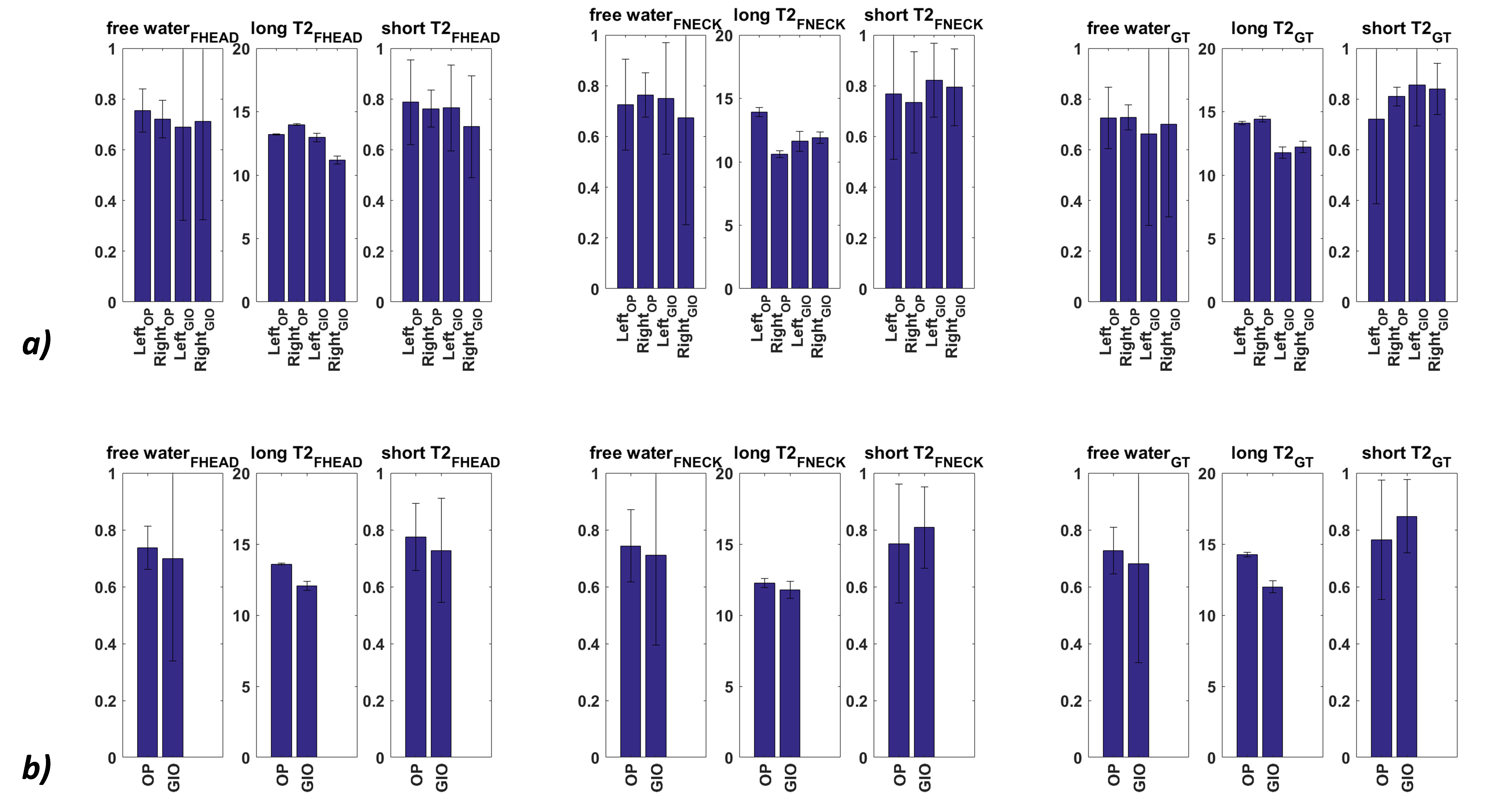
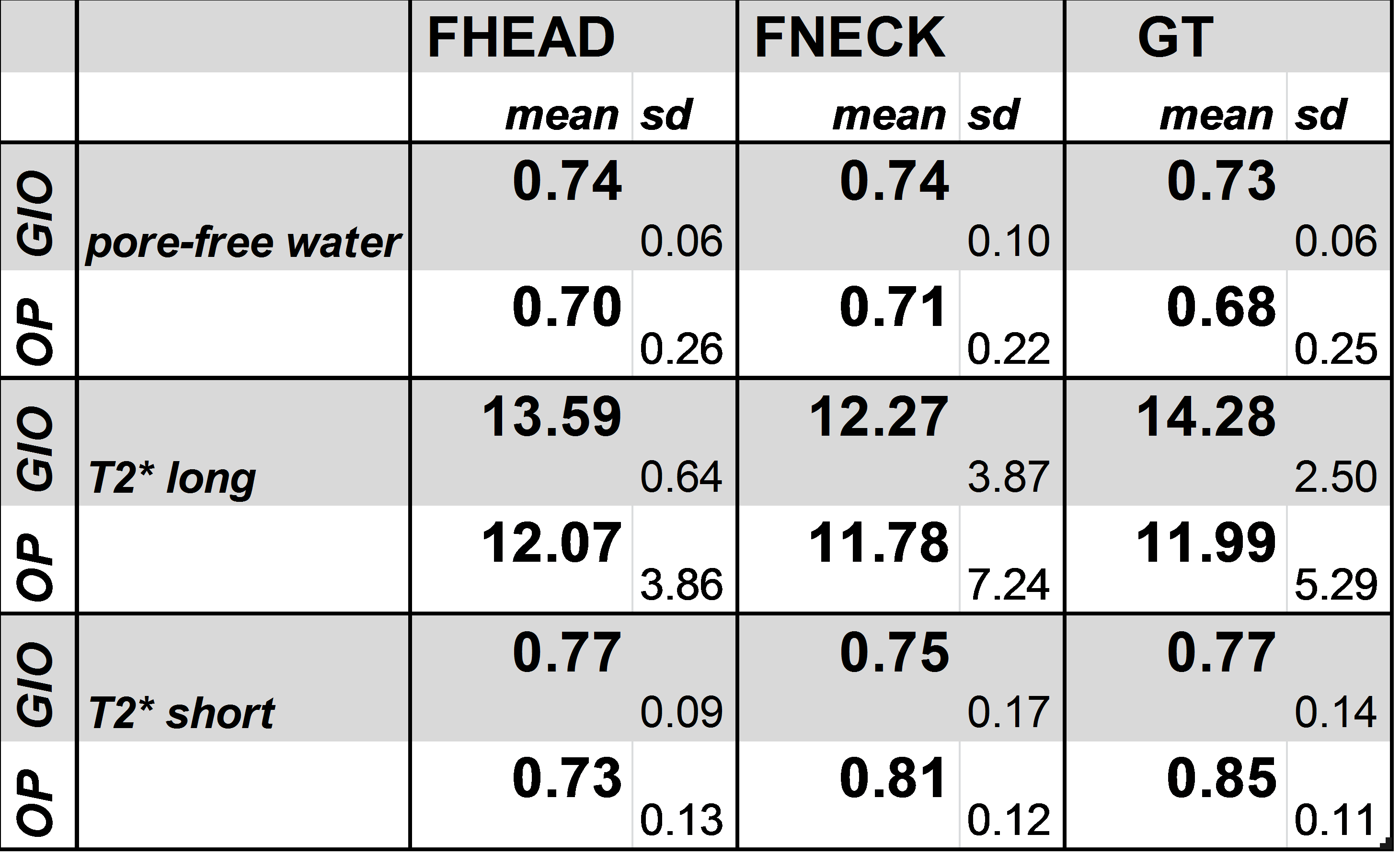
This study had institutional review board approval and written informed consent was obtained. MRI acquisition was performed at 3T using an uTE Spiral VIBE sequence6,7 on n=9 subjects (4 OP subjects: BMI=27.2+/-3.9kg/m^2 , age =54+/-2y; and 5 GIO subjects: BMI =22.1+/-12.9 kg/m^2 , age =34+/-12y ). Two sets of one-shot, 6-echoes uTE-VIBE data were acquired with following echo times: (1) 0.080- 2.4-4.8-7.29.6-12-14.4ms / (2) 0.200- 3.6 – 6.0 – 8.4 – 10.8 – 13.2 – 15.6ms and the following parameters: TR= 16ms, NA= 1, 48 axial slices, In-plane resolution = 1.4x1.4mm, Slice thickness = 2.3mm. 3D registration was performed using rigid transformation to avoid displacement between the two sets of image. 24 axial slices were chosen in the proximal femur to cover following ROIs: Femoral Head (fNeck), Neck (fNeck) and Great Trochanter. Based on the 0.080 ms uTE images, cortical bone was segmented in these subregions using in-house MATLAB script. Using a non-linear least square fitting procedure, ROIs were fitted pixel-by-pixel according to a bi-exponential model (Fig1a) to obtain pore-free water fraction, T2*short, T2*long in Left and Right Hip.Results
Typical uTE acquired images and parametric maps in segmented ROIs are shown respectively in Fig1b and Fig1c. Figure 2 shows the average value in each ROI in the left and right hips for GIO and OP subjects. No significant differences were found within subjects between the left and right hips and between GIO and OP subjects. Values of T2*short (0.7-0.8ms) and T2*long (12-14ms) (Table1) were similar to those previously reported in proximal femur3. Pore-free water fraction was elevated compared to previously reported values in the literature (~0.7 compared to ~0.4), but this may be because this is an in vivo as opposed to specimen study and because this is a study of subjects with osteoporosis, in which higher porosity of cortical bone and thus pore water is expected. Globally, pore water was not significantly different between GIO and OP subjects. T2* long tended to be longer in subregions for GIO compared to OP subjects. Within GIO and OP subjects, there were subregional differences in T2* long. The mean T2* short of all subregions was 0.76ms in GIO subjects and 0.79 ms in OP subjects. Within OP subjects, there were also subregional differences in T2* short. [GC1] [a2] [GC1]Were these differences statistically significant? [a2]No statistical difference at this point mainly due to the high sd for the 4 OP subject…Conclusion
In this preliminary study, there were no significant differences in cortical bone porosity indicated by uTE parameters in GIO compared to OP subjects. In both GIO and OP subjects, there was subregional variation in the proximal femur in T2* long and T2* short. This may reflect subregional differences in bone quality of the cortex of the proximal femur. More subjects are needed to confirm these results.Acknowledgements
No acknowledgement found.References
1. Juras V, Apprich S, Zbýň Š, et al. Quantitative MRI Analysis of Menisci Using Biexponential T(2)* Fitting with a Variable Echo Time Sequence. Magnetic resonance in medicine : official journal of the Society of Magnetic Resonance in Medicine / Society of Magnetic Resonance in Medicine. 2014;71(3):1015-1023.
2. Ma Y-J, Chang EY, Bydder GM, Du J. Can Ultrashort Echo Time (UTE) Magnetic Resonance Imaging Sequences on a 3T Clinical Scanner Directly Detect Signal from Collagen Protons: Freeze-Dry and D(2)O Exchange Studies of Cortical Bone and Achilles Tendon Specimens. NMR Biomed. 2016;29(7):912-917.
3. Nazaran A, Carl M, Ma Y, et al. Three-dimensional adiabatic inversion recovery prepared ultrashort echo time cones (3D IR-UTE-Cones) imaging of cortical bone in the hip. Magn Reson Imaging. 2017;44:60-64.
4. Bae WC, Chen PC, Chung CB, Masuda K, D’Lima D, Du J. Quantitative Ultrashort Echo Time (UTE) MRI of Human Cortical Bone: Correlation with Porosity and Biomechanical Properties. Journal of bone and mineral research : the official journal of the American Society for Bone and Mineral Research. 2012;27(4):848-857.
5. Kageyama G, Okano T, Yamamoto Y, et al. Very high frequency of fragility fractures associated with high-dose glucocorticoids in postmenopausal women: A retrospective study. Bone Reports. 2017;6(Supplement C):3-8.
6. Grodzki DM, Jakob PM, Heismann B. Ultrashort echo time imaging using pointwise encoding time reduction with radial acquisition (PETRA). Magn Reson Med. 2012;67(2):510-518.
7. Qian Y, Boada FE. Acquisition-weighted stack of spirals for fast high-resolution three-dimensional ultra-short echo time MR imaging. Magn Reson Med. 2008;60(1):135-145.
Figures