5192
SyntheticTSE: Accelerated multicontrast spin-echo knee MRI using a combination of Bloch-simulation models, compressed sensing and simultaneous multislice acquisition1Center for Advanced Imaging Innovation and Research (CAI2R), NYU School Of Medicine, New York, NY, United States, 2Biomedical Engineering, School of Engineering, New York University, Brooklyn, NY, United States, 3Department of Biomedical Engineering, Tel Aviv University, Tel Aviv, Israel, 4Sagol School of Neuroscience, Tel Aviv University, Tel Aviv, Israel, 5Department of Radiology, NYU School Of Medicine, New York, NY, United States, 6Center for Biomedical Imaging, Department of Radiology, NYU School Of Medicine, New York, NY, United States
Synopsis
Clinical knee MRI examinations usually consist of a series of 2D acquisitions with different contrasts and orientations. In this work, we propose to simplify and shorten the examination by performing multi-contrast imaging using a single acquisition by directly reconstructing parametric maps (PD and T2) and then synthesizing the contrasts of clinical interest (PD-weighted and T2-weighted with and without fat suppression). The proposed SyntheticTSE method uses a Bloch-simulation signal model for parametric mapping and a combination of compressed sensing and simultaneous multislice acceleration. Initial feasibility is demonstrated using retrospective undersampling of multi-spin-echo knee dataset
Purpose
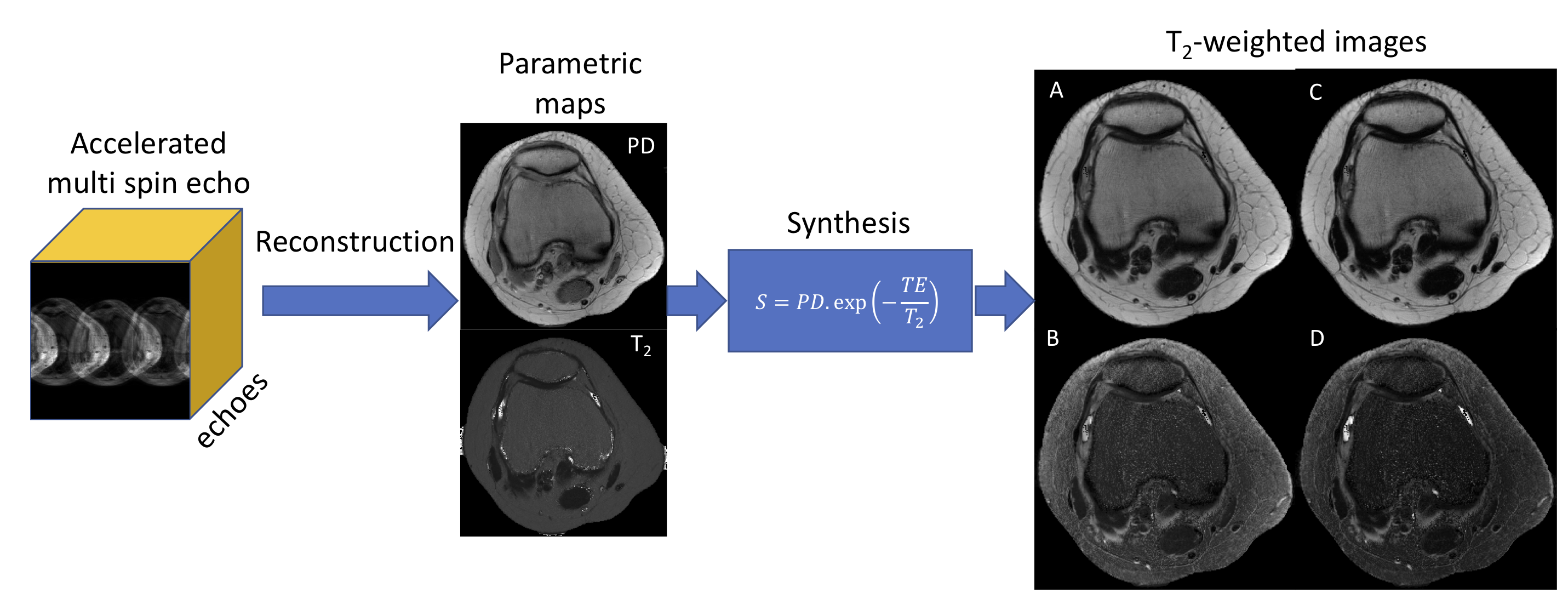
Clinical knee MRI protocols typically include multiple 2D acquisitions with different contrasts and orientations, resulting in long examinations times. Compressed sensing (CS) [1], simultaneous multislice (SMS) [2] and combinations of these reconstruction techniques [2,3] can reduce the time of individual acquisitions, but do not reduce the need for multiple acquisitions. In addition, each acquisition is vulnerable to T2-blurring from long echo trains. Recent developments in quantitative imaging, such as fingerprinting [4] and Bloch-simulation-based T2 mapping [5,6], enable the reconstruction of multiple parametric maps, such as PD, T1 and T2, from a single acquisition. In this work, we present a rapid spin-echo technique named SyntheticTSE that can directly reconstruct PD and T2 maps and synthesize arbitrary clinical contrast for knee MRI from an accelerated single multi spin-echo (MSE) acquisition using a combination of Bloch-simulation signal modeling, CS and SMS (Fig. 1).Methods
A 2D MSE acquisition accelerated with in-plane CS (random ky undersampling) and through-plane SMS was employed to simultaneously encode PD, T2 and transmit field distribution (B1+). The relationship between these physical parameters are modeled using the echo-modulation curve (EMC) approach, as proposed in [5]:
$$ {\psi(\rho,T_2,B_1)=EMC(T_2,B{_1}{^+})\cdot{C}.\cdot{\rho} \space{...}\space (1)} $$
Using this forward model, PD, T2, and B1+ maps are directly computed from k-space data by solving the inverse problem which minimizes the following cost function:
$$ {\phi(\rho,T_2,B{_1}{^+})=\frac{1}{2}||F\cdot{\psi}-y||^2_2+\lambda_\rho||W_\rho\cdot\rho||_1+\lambda_{T_{2}}||W_{T_{2}}\cdot{T_2}||_1+\lambda_{B_{1}}||W_{B_{1}}\cdot{B{_1}{^+}}||_{1}\space{...}\space{(2)}} $$
where $$$y$$$ is the undersampled MSE k-space data, $$${\psi}$$$ is the EMC operator that models PD, T2 and B1, $$$F$$$ is the Fourier transform according to the sampling pattern, $$$\lambda_i$$$’s are regularization factors and $$$W_i$$$’s are regularization transforms (spatial Wavelets to exploit sparsity in the parametric maps). Images with synthetic contrast are generated using PD and T2 maps for different echo times (TE):
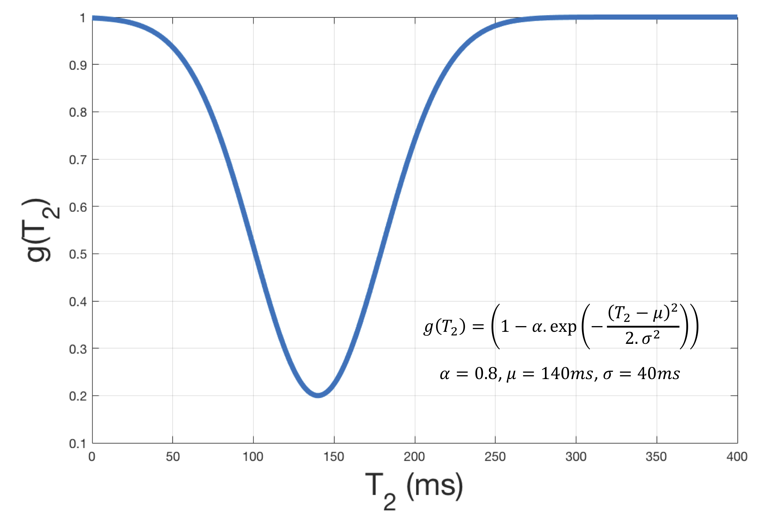
$$ {S=PD \cdot \exp{-\frac{TE}{g(T_2)}} \space{...}\space (3)} $$
where $$$ g(T_2) $$$ performs fat suppression by attenuating regions with a T2 to the ones reported for fat tissue [7] (see Fig. 2 for more details).
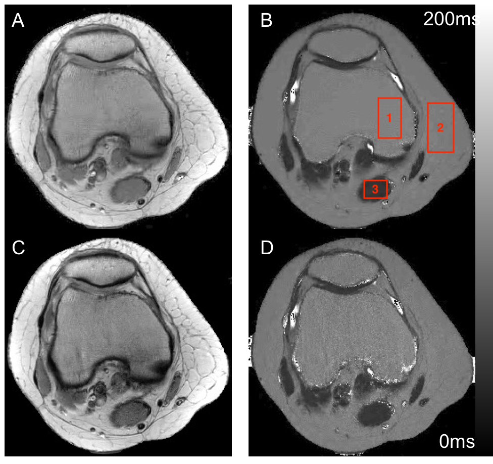
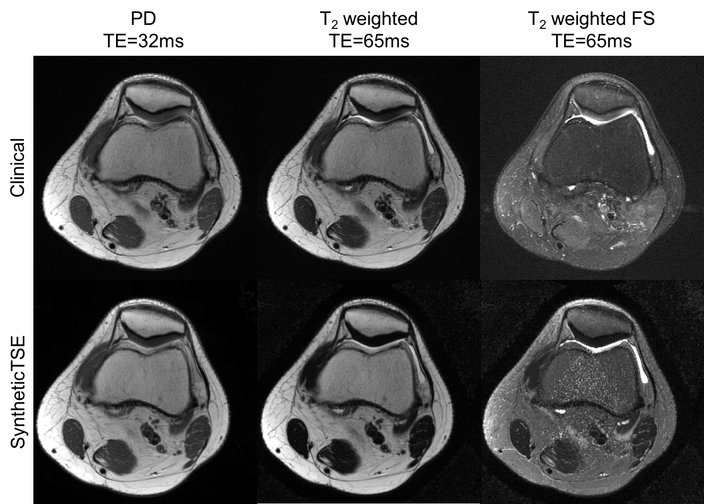
SyntheticTSE was evaluated on a fully-sampled MSE knee data acquired on axial orientation using a 3T Siemens Skyra scanner with a 15-channel coil array. Relevant imaging parameters include: image matrix = 256x256, 6 slices, FOV = 140x140mm2, TR = 3.5s, TE = 10ms, 20 echoes, slice thickness = 3mm, scan time = 15 min. The acquired dataset was retrospectively undersampled in-plane by CS factors 2, 3 and 4, and through-plane by SMS factors 2, 3 and 4. Maximum acceleration was selected based on the PD and T2 error between the accelerated and fully-sampled reconstructions. Synthetic TSE results with PD, T2 and fat-saturated T2 were compared against three different conventional TSE acquisitions. The total scan time for the conventional acquisitions was 6:48 min.
Results
The combined CS and SMS approach can accelerate the acquisition of MSE up to a factor of 6 (2-fold in-plane CS and 3-fold SMS accelerations), resulting in PD and T2 maps with less than 7% and 5% averaged error respectively (Fig. 3). T2 tissue values were also in concordance with values listed in previous work [7], 122.9±4.6ms (ROI 1, bone marrow), 123.3±4.6ms (ROI 2, fat) and 35.3±1.6ms (ROI 3, muscle). Synthesized T2-weighted images without fat suppression achieved similar quality and contrast as conventional TSE acquisitions (Figure 4). Fat-suppressed (FS) images also presented a contrast close to conventional TSE, at the expense of SMS-related noise amplification at the center.Discussion
SyntheticTSE proposes to synthesize contrast by reconstructing MR parameter maps directly from a single acquisition instead of using different acquisitions with fixed contrast. The proposed method provides stable and reproducible contrast for different scan settings by using ideas from model-based reconstruction and fingerprinting [8], in addition, acceleration based on CS and SMS reduces the acquisition time making it feasible for clinical studies. Compared to other multicontrast knee MRI approaches such as T2-shuffling [9], SyntheticTSE also offers access to quantitative information provided by the reconstructed PD and T2 maps.
Conclusion
The combination of Bloch simulation-based modeling of the temporal signal evolution in multi-spin-echo imaging and acceleration using CS and SMS enables to perform multicontrast MRI without and with fat suppression from a single acquisition. The proposed SyntheticTSE can generate flexible image contrast and enable access to quantitative T2 information, potentially reducing the number of acquisitions in the conventional knee MRI protocol and therefore the time required for imaging.
Acknowledgements
Funding: 1P41EB017183-01A1References
1. Recht M, Otazo R, Rybak L, Gyftopoulos S, Petchprapa C, Geppert C, Bruno M, Raithel E. 3D TSE imaging using SPARSE-SENSE acceleration: Comparison with conventional 2D TSE imaging for detection of internal derangement of the knee. Proceedings ISMRM 2015, Toronto, Canada, p. 1198.
2. Fritz J, Fritz B, Zhang J, Thawait GK, Joshi DH, Pan L, Wang D. Simultaneous Multislice Accelerated Turbo Spin Echo Magnetic Resonance Imaging: Comparison and Combination With In-Plane Parallel Imaging Acceleration for High-Resolution Magnetic Resonance Imaging of the Knee. Invest Radiol. 2017 Sep;52(9):529-537.
3. Yoshimoto A, Baete S, Bruno M, Kline M, Garwood E, Boada F, Otazo R, Recht M. Rapid knee MRI using TSE sequences accelerated with a combination of simultaneous multislice, multicoil compressed sensing and elongated echo trains. Proceedings ISMRM 2017, Honolulu, p. 1150.
4. Ma D, Gulani V, Seiberlich N, Liu K, Sunshine J, Duerk J, Griswold M. Magnetic resonance fingerprinting. Nature 2013;14:187–192.
5. Ben-Eliezer N, Sodickson DK, Shepherd T, Wiggins GC, Block KT. Accelerated and motionrobust in vivo T2 mapping from radially undersampled data using bloch-simulation-based iterative reconstruction. Magn Reson Med 2016; 75(3): 1346-54.
6. Ben-Eliezer N, Sodickson DK, Shepherd T, Wiggins GC, Block KT. Accelerated and motionrobust in vivo T2 mapping from radially undersampled data using bloch-simulation-based iterative reconstruction. Magn Reson Med 2016; 75(3): 1346-54
7. Pai, A, Xiaojuan Li, and Sharmila M. “A Comparative Study at 3 Tesla of Sequence Dependence of T2 Quantitation in the Knee.” Magnetic resonance imaging 26.9 (2008): 1215–1220. PMC. Web. 2 Nov. 2017.
8. Shepherd TM, Kirov II, Charlson E, Bruno M, Babb J, Sodickson DK, Ben-Eliezer N. New rapid, accurate T 2 quantification detects pathology in normal-appearing brain regions of relapsing-remitting MS patients. NeuroImage: Clinical. 2017 Dec 31;14:363-70.
9. Tamir, J I., et al. "T2 shuffling: Sharp, multicontrast, volumetric fast spin‐echo imaging." Magnetic resonance in medicine 77.1 (2017): 180-195.
Figures