5190
New Tissue Saturation Method for the Detection of Therapeutic Cells in a Knee JointDjaudat Idiyatullin1, Michael Garwood1, and Sergey Magnitsky2
1Center for Magnetic Resonance Research and Department of Radiology, University of Minnesota Medical School, Minneapolis, MN, United States, 2Radiology, UCSF, San Francisco, CA, United States
Synopsis
Synapsis. Mesenchymal Stem Cells (MSCs) have a high potential for a treatment of bone diseases. We develop new acquisition protocol for the detection of therapeutic cells in knee joints. Labeling MSCs with iron oxide particles not only reduce T2* but also induce the resonance frequency shift of labeled cells. This shift enabled us to implement tissue saturation scheme and detect distinct hyperintense signal from grafted cells. New protocol allowed us to detect and quantify therapeutic cells for six days after implantation which was not possible before. Proposed protocol opens new opportunities for in vivo monitoring of cell therapy of bone disorders.
Introduction
It has been shown histologically that Mesenchymal Stem Cells (MSCs) have high potentials for a treatment of bone loss diseases such as osteoporosis, osteoarthritis and bone fracture1,2. An injection of MSCs directly into a knee joint has been suggested, as an effective delivery method of the therapeutic cells. However, the fate of the grafted cells after administration is unknown until histological staining. Our group is developing noninvasive techniques to detect and quantify grafted cells overtime and to facilitate the translation of promising preclinical results into the clinical. Conventional T2-weighted MRI method was successfully implemented for the detection of iron labeled cells in brain, spine and muscle tissues. However, this technique produces a hypointense signal from both bones and grafted cells, which make the detection and quantification of the therapeutic cells very challenging in musculoskeletal systems. Our previous studies indicate that the SWIFT3 pulse sequence enables to produce a hyperintense signal from iron labeled cells4. In this project, we modified original SWIFT acquisition protocol and implemented new magnetization preparation scheme. This scheme suppressed signal from the host tissue and significantly improved in vivo detection of the grafted cells.Methods
Cell culture: Mouse MSCs were maintained on uncoated flasks in media with DMEM and 10% FBS and labeled with different concentration of Feraheme overnight5. Phantoms: To minimize bulk magnetic susceptibility gradients spherical NMR tubes with different iron oxide solutions were immersed into cylindrical NMR tube with pure water and a chemical shift of the iron solutions relative to pure water signal was measured with a pulse-acquire sequence. All spectra and images were acquired at 9.4T animal scanner (Agilent Technologies, USA). MSCs injection: 20 ml of iron-oxide labeled MSCs (200 μg/ml of Fe) were injected into the knee joint of rats’ highlimbs. Imaging: Radial 3D MBSWIFT images6 were acquired with 4-phase shifted hard pulses of 5.2 μs length (with 2.6 μs dead time), TR = 1.9 ms, excitation/acquisition bandwidths 192/384 kHz, number of views = 96000. Frequency selective RF pulse (Gaussian, 7 ms, 90-degree flip angle) tuned to the water proton signal was implemented for a tissue saturation.Results
We discovered that the iron oxide particles not only reduced the T2 relaxation time but also increased resonance frequency of surrounding water molecules. NMR spectrum of an iron solution in a spherical NMR tube and pure water exhibited two distinct signals (Fig.1A). The peak on the left is the signal from iron-oxide solution while peak on the right is from pure water. The shift of the proton frequency in the iron solutions was linearly proportional to the concentration of the iron oxide particles (Fig1.B) This frequency shift opens an opportunity for us to saturate water signal from the tissue without significant changes of the signal from iron labeled cells. Fig.2C shows anatomical SWIFT image of rat knee joint, D – depicts this knee after application of tissue saturation scheme, E – tissue saturated SWIFT image of the knee joint after administration of 4 x10⁶ iron labeled MSC. Hypointense signal from grafted cells was clearly detected in these experiments. New imaging protocol allowed us to detect and quantify grafted cells for six days after the administration.Discussion
High concentration of labeling contrast agent is very beneficial for the detection of grafted cells. It provides stronger contrast and longer detection time of the injected cells. In this study, we discovered that iron oxide particles induced the chemical shift of protons signal from water molecules, which became very significant at a high concentration of the contrast agent. The resonance frequency shift produced by iron oxide particles allowed us to implement an additional RF pulse and suppress the NMR signal from the normal tissue while the signal from iron labeled cells remained unaffected. With our new method, we were able to detect and quantify grafted MSC for six days after administration, which was not possible before. Our new imaging protocol opens earlier not available opportunities to monitor stem cell therapy in complex anatomical areas such as knee joints over time. Our method should be applicable to any cell tracking studies and can find many applications for stem- and immuno-therapies.Acknowledgements
This study was supported by NIH grants R21A06850 and P41 EB015894. We also would like to thank Tony Huynh for the help with MRI acquisition and animal preparations.References
- Guan M, Yao W, Liu R, et al. Directing mesenchymal stem cells to bone to augment bone formation and increase bone mass. Nature medicine 2012;18:456-62.
- Yao W, Guan M, Jia J, et al. Reversing Bone Loss by Directing Mesenchymal Stem Cells to the Bone. Stem Cells 2013.
- Idiyatullin D, Corum C, Park JY, et al. Fast and quiet MRI using a swept radiofrequency. J Magn Reson 2006;181:342-9.
- Magnitsky S, Idiyatullin D, Corum C, et al. Imaging of Grafted Mesenchymal Stem Cells in Bone Tissue. Proc. Intl.Soc.Mag.Reson.Med. 2015;15:1020.
- Frank JA, Zywicke H, Jordan EK, et al. Magnetic intracellular labeling of mammalian cells by combining (FDA-approved) superparamagnetic iron oxide MR contrast agents and commonly used transfection agents. Academic Radiology. 2002;9:S484-7.
- Idiyatullin D, Corum CA, Garwood M. Multi-Band-SWIFT. J Magn Reson 2015;251:19-25.
Figures
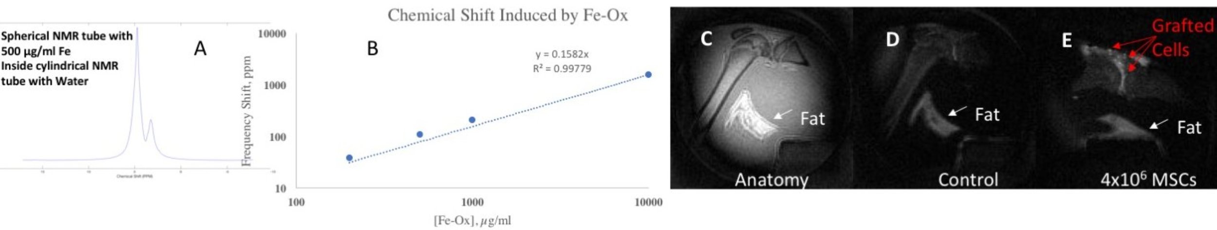
A-NMR spectrum of iron oxide solution and waters in two
different compartments. The iron oxide solution has different resonance
frequency than pure water. B-Linear dependence of the frequency shift of the
proton signal in the iron oxide solution on the iron oxide concentration.
C-SWIFT image of a rat knee joint, D-tissue suppressed SWIFT image of the same
knee joint, E-tissue suppressed SWIFT image of the same knee joint after
administration of iron-labeled MSCs