5186
Novel MR coil hardware and fast 3D imaging sequence in evaluating ankle kinematic instability1Radiology Department of Peking University Third Hospital, Beijing, China, 2United Imaging Healthcare, Shanghai, China
Synopsis
A specially designed Ankle & Foot coil that adapts Achilles Tendon Boot is utilized to multiple angle imaging of ankle kinematic instability. With the 3D fast spin echo sequence with compressed sensing based acceleration technique, multi angle high resolution ankle imaging can be achieved in less than 20 minutes. Images from the volunteer with a history of ankle injuries showing anterior talar translation and rotated internally. The integration of novel coil and compressed sensing based acceleration technique enabled ankle kinematic imaging into clinical routine.
Introduction
Ankle instability frequently occurs after the injury of lateral collateral ligament (LCL). Due to the limitation of SNR, temporal and spatial resolution, MR scan is designed to detect ligament injury rather than the joint instability in the diagnosis of ankle instability. Ankle is also fixed at a neutral position by the coil during MRI examination to avoid possible motion artifact, which precludes the analysis of ankle stability in normal ankle movement. We developed a specially designed Ankle & Foot coil that adapts Achilles Tendon Boot and high resolution sequence to provide a direct observation of the ankle kinematical stability. Methods
Methods
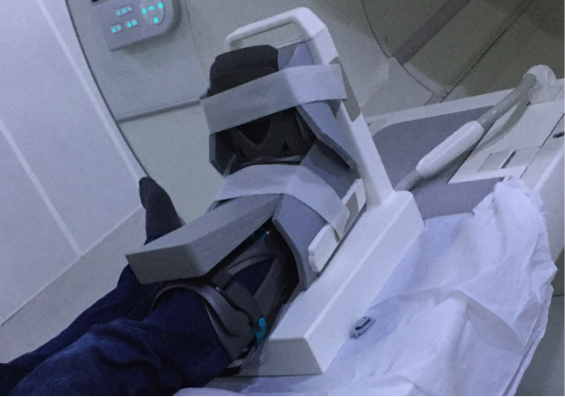
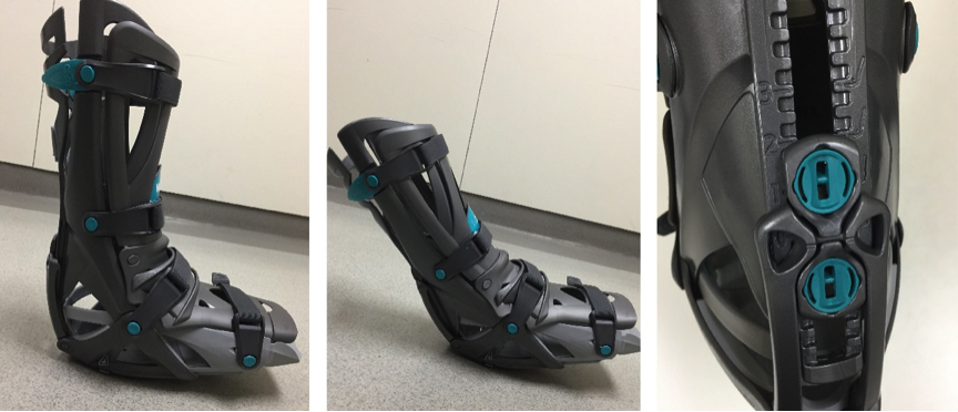
We developed a 24-channel receive-only coil array customized for foot and ankle imaging. The coil includes four wing-shaped pads in flexible material to better conform to ankle. It is open upside and is able to hold an Achilles Tendon Boot. 14 loops around the ankle provide enough SNR and acceleration potential for ankle imaging (Figure 1).
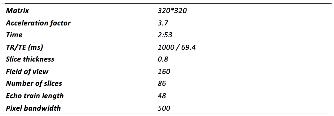
With the help of Achilles Tendon Boot in the coil, the ankle can be firmly fixed in more than twenty angles (Figure2). Five angulations were manipulated from maximal plantar flexion to maximal dorsal flexion. For each position of the ankle, a 3D PDWI fast spin echo sequence with variable flip angle (sequence parameters are listed in table 1) is used to evaluate the ankle. 0.5mm*0.5mm*0.8mm voxel images were acquired in less than 3minutes with compressed sensing based acceleration technique. Image data were acquired on a 3T system ( uMR 770, UIH, Shanghai, China).
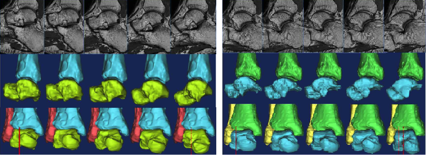
To trace the movement process of the talus, the registration of tibia is implemented. Then images from five different angles are gathered and reviewed by an experienced radiologist in a viewer (Mimics 15.0 of Materialis). Two volunteers were included in the present study. Informed content was obtained from all volunteers in accordance with the institutional review board policy. One volunteer has no history of ankle injury. The other has recurrent ankle sprains and is diagnosed with a rupture of anterior talus fibular ligament.
Results
The Achilles Tendon Boot offers good fixation of ankle in different angles. High quality Images without motion artifact were thus acquired. The 3D fast spin echo sequence provides a great delineation of the anatomical structures in ankle (Figure 3).
Images from the volunteer with a history of ankle injuries had anterior talar translation and internal rotation. Posterior part of the ankle joint space was widened and anterior part of the joint space was narrowed. In terms of images from the control volunteer, no talus rotation was observed. And the joint space was always constant in various angles of ankle.
Discussion
The function of LCL is to keep the talus from anterior translation and internal rotation 1. In this investigation, talus rotation and joint space variation is observed in the patient with a history of ankle injuries during ankle movement. This is a clear and objective evidence for diagnosis of ankle instability 2. In addition, the narrowing of the anterior joint space may cause anterior impingement of ankle and lead to osteochondral lesion of talus. This could also help predict possible lesions in patients with ankle instability.
Some previous studies reported that ankle instability evaluation can be fulfilled with multiple angle CT scan or roentgen stereo photogrammetry 3,4. However, given the ionizing radiation and relative low soft tissue contrast of CT scan, MRI seems to be a better choice for imaging muscular skeletal injury. Ankle instability evaluation requires high resolution and multiple angle imaging of the ankle joint, which raises challenges to MRI hardware and software. The new multi-channel coil provides great SNR for high resolution MRI imaging of ankle anatomy. The Achilles tendon boot offers stable and comfortable fixation of the ankle in different angles, which enables the kinematical analysis of ankle stability. The 3D fast spin echo sequence clearly revealed the ankle structures. All image acquisition and reconstruction is finished at the scanner in a short time.
Conclusion
With a specially designed Ankle & Foot coil that adapts Achilles Tendon Boot, multiple angle imaging of ankle kinematic instability can be fulfilled. With the 3D fast spin echo sequence with compressed sensing based acceleration technique, multi angle high resolution ankle imaging can be achieved in less than 20 minutes. The integration of novel coil and compressed sensing based acceleration technique enabled ankle kinematic imaging into clinical routine.Acknowledgements
No acknowledgement found.References
1. Wikstrom E A, Hubbard-Turner T, Mckeon P O. Understanding and Treating Lateral Ankle Sprains and their Consequences[J]. Sports Medicine, 2013, 43(6):385-393.
2. Leardini A, O'Connor J J, Catani F, et al. Kinematics of the human ankle complex in passive flexion; a single degree of freedom system[J]. Journal of Biomechanics, 1999, 32(2):111-118.
3. Lundberg A, Goldie I, Kalin B, et al. Kinematics of the ankle/foot complex: plantarflexion and dorsiflexion[J]. Foot & Ankle, 1989, 9(4):194.
4. Wang B, Roach K E, Kapron A L, et al. Accuracy and feasibility of high-speed dual fluoroscopy and model-based tracking to measure in vivo ankle arthrokinematics.[J]. Gait & Posture, 2015, 41(4):888-893.
Figures