5183
Comparison of ZTE vs UTE vs Cones for Ultrashort TE MSK Imaging1GE Healthcare, San Diego, CA, United States, 2UCSD, San Diego, CA, United States
Synopsis
We compared different center out 3D radial trajectories and assess their advantages and disadvantages for short T2 MSK imaging. We found that while ZTE and Cones may provide some unique capabilities, UTE represents a good compromise between the two.
Introduction
Direct MR imaging of tissues such as tendons, or ligaments, which have short transverse relaxation times (T2s) has become possible using ultrashort echo time (UTE) sequences [1-4]. There are several excitation RF and k-space trajectories available for UTE imaging. In this work, we compare different center out 3D radial trajectories and assess their advantages and disadvantages for short T2 MSK imaging.Theory
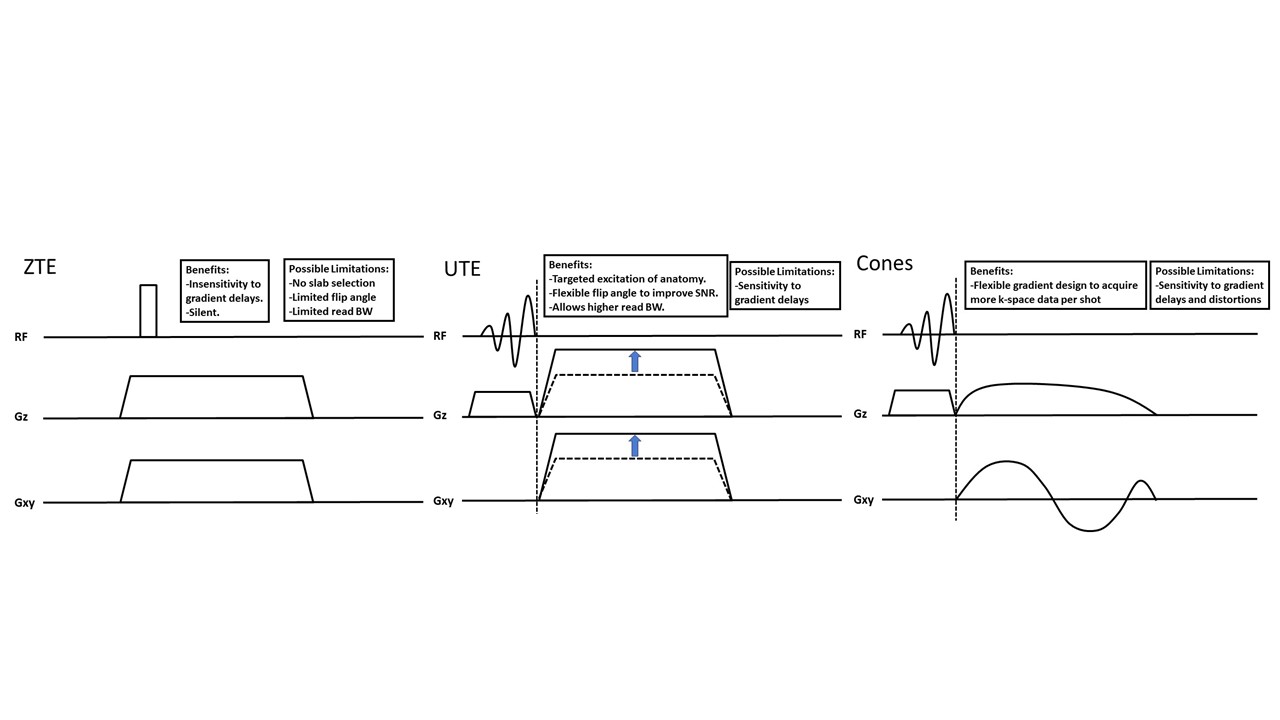
The pulse sequences are shown in Fig.1. Both UTE and Cones employ their RF pulses while the read gradients are ramped down, while in ZTE the read gradients are already ramped-up during excitation (see Fig.1A). Some consequences are:
1) The flip angle in ZTE is limited due to the need to keep the RF excitation pulse very short.
2) UTE and Cones can apply a slice-selection gradient, which allows application of slab selection.
3) UTE and Cones acquire data during partially time-varying read gradients, while ZTE uses an already fully ramped read gradient which means that ZTE does not suffer from gradient delay errors.
4) Both UTE and Cones allow to acquire multiple echoes at later TEs.
5) Cones has a flexible read gradient design that allows a spiral-like k-space trajectory, which can cover more k-space data per RF excitation.
Methods
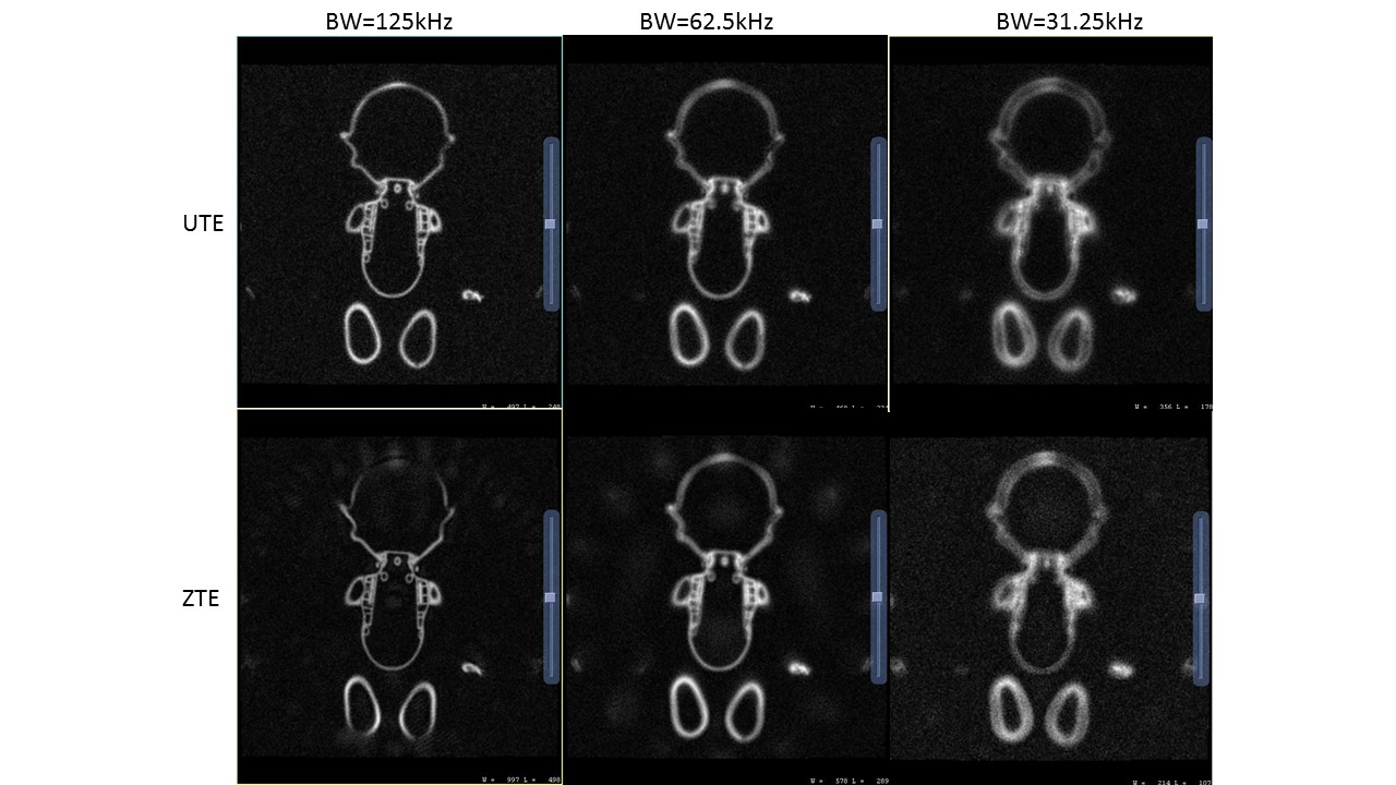
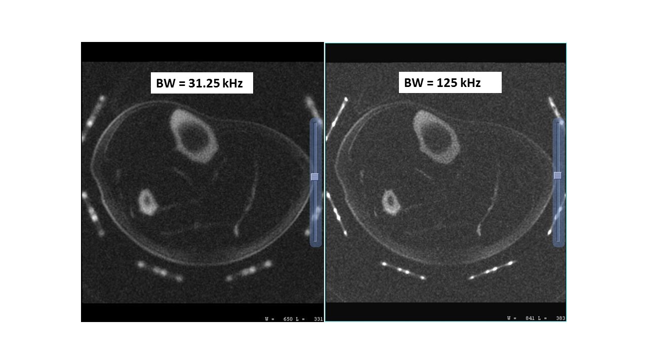
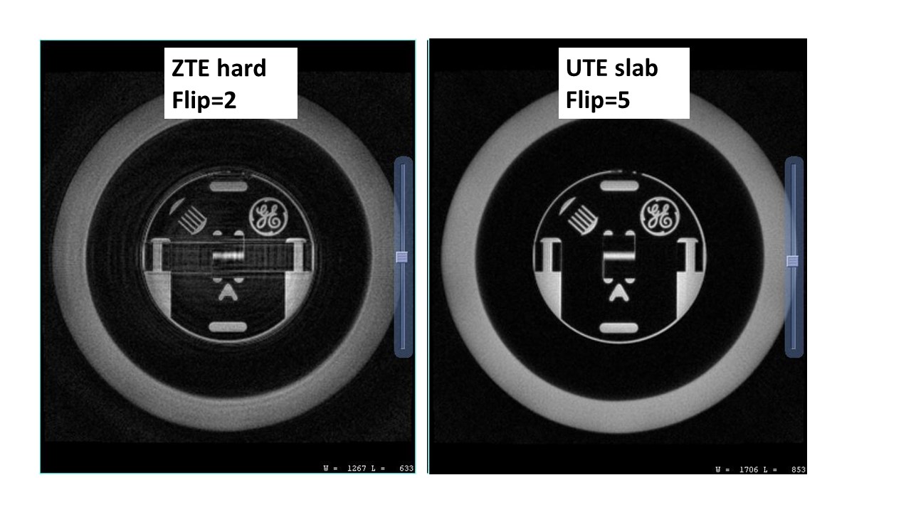
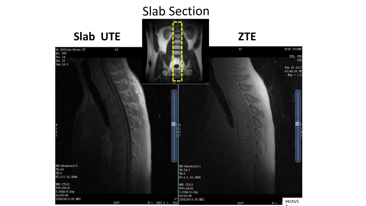
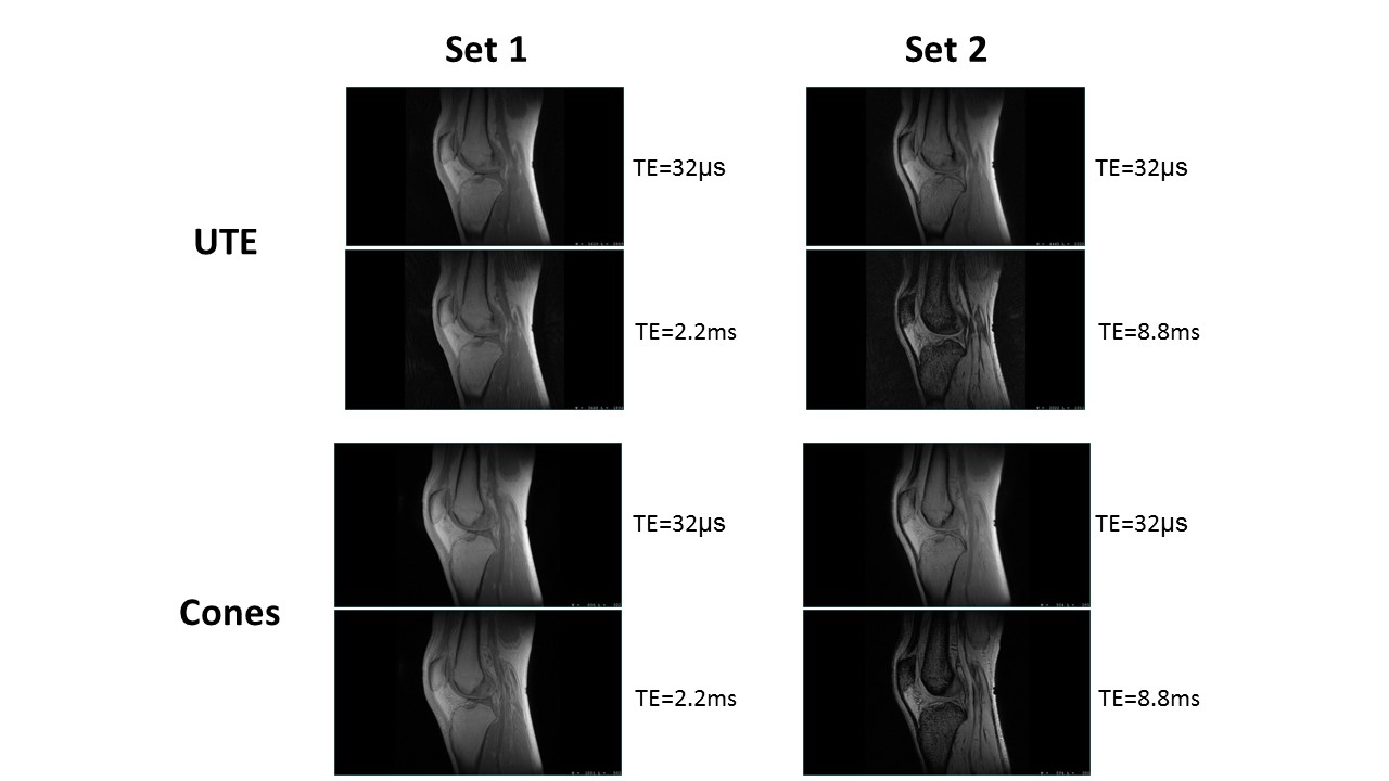
To experimentally study the blurring characteristics, we scanned a plastic doll made of soft rubber (short T2* of about 400𝜇s). Both UTE and ZTE scans were performed with BW of 31.25kHz, 62.5kHz, and 125kHz. For the in-vivo scans we imaged the axial tibia using inversion recovery (IR) prepared UTE at two BWs (31.25kHz and 125kHz). To study the RF excitation, we scanned a resolution-phantom in the axial plane, focusing only on a small axial section that contained structure. The UTE scan were acquired using slab selective pulses (at Ernst angle) to show the best possible image quality (artifacts and SNR). Similar scans were performed in the sagittal spine. Finally, multi-echo UTE and Cones scans were performed in the sagittal knee. Two separate dual echoes acquisitions were obtained to highlight the flexibility of the Cones trajectory. Relevant scan parameters were FOV=25cm, matrix=256, slice thickness=3mm, 20slices, BW=125kHz. For the first scan, the second echo was chosen near the minimum available echo spacing (TE2=2.2ms). For the second acquisition, the second echo was chosen larger at TE2=8.8ms, meaning that the Cones sequence was able to acquire more k-space data per TR period.Results
The images of the short T2 doll-phantom are shown in Fig.2. For either sequence there is significant short T2 blurring for the 31.25kHz BW scans, while the best result is obtained with UTE using BW=125kHz. The axial tibia images are shown in Fig.3, which show similar blurring for the lower BW acquisitions. The axial scans in the resolution phantom to study the RF excitation differences are shown in Fig.4. Fig.4A shows the center slice of the ZTE acquisition. Since no slab selection was applied, there exist a lot of excited signals from the phantom outside the encoded 3D volume, which manifests as artifacts in the final image. Fig.4B shows the UTE acquisition using a slab-selective RF pulse. The corresponding in-vivo images are shown in Fig.5 in the sagittal spine. Since no slab-selection was applied in ZTE, there exists a lot of excited signals from either the left or right side of the spine which manifests as artifacts in the final image. In the corresponding UTE acquisition using a slab-selective RF pulse, these artifacts have been eliminated. Finally, Fig.6 shows several dual-echo scans using UTE (top) and Cones (bottom). The left column corresponds to the acquisitions with the shorter second echo (TE2=2.2ms). Here both UTE and Cones show similar image quality and SNR. The right column shows the corresponding results for the longer second echo (TE2=8.8ms). In this case, compared to UTE, the Cones acquisition shows both less artifacts and more SNR, due to the more efficient k-space sampling duty cycle of Cones.Conclusion
We have examined three ultra-short-TE pulse sequences for their performance in SNR, artifact characteristics and multi-echo capability. While ZTE and Cones may provide some unique capabilities, UTE represents a good compromise between the two. Although our current version of ZTE was not able to acquire multi-echo images, future work may include a version of that psd that has such capability (such as looping-star).Acknowledgements
No acknowledgement found.References
[1] Rahmer et al, Magn Reson Med, 2006. 55(5): p.1075-82.
[2] Du et al, Magn Reson Imag 2011:29:470–482
[3] Weiger et al, NMR Biomed. 2015 28(2):247-54
[4] Li et al, Magn Reson Med 2012 68(3):680
Figures