5175
Water/fat separation imaging at 7T using 3D integrated SSFP1State Key Laboratory of Brain and Cognitive Science, Beijing MRI Center for Brain Research, Institute of Biophysics, Chinese Academy of Sciences, Beijing, China, 2University of Chinese Academy of Sciences, Beijing, China, 3Siemens Shenzhen Magnetic Resonance Ltd, Shenzhen, China, 4Stevens Neuroimaging and Informatics Institute, University of Southern California, Los Angeles, CA, United States
Synopsis
Balanced SSFP (bSSFP) has been used as a new alternative for water-fat separation imaging due to its fast acquisition and high SNR efficiency. However, it displays characteristic banding artifacts in the presence of field inhomogeneity, especially at the 7T ultrahigh-field MRI system. Complex combination of two phase-cycled bSSFP was used to alleviate the artifact at 3T, but would take more scan time. Integrated SSFP (iSSFP), which was modified from bSSFP, was introduced to separate water and fat while removing banding artifacts in shorter scan time. iSSFP also inherited the advantages of relatively high SNR and bright fluid. 3D iSSFP with high spatial resolution may have great potential to separate water and fat in the clinical application of 7T ultrahigh-field MRI.
Introduction
A variety of water-fat separation methods, including fast spin-echo (FSE) and gradient recalled echo (GRE), were utilized to improve visualization of abnormalities. In the past decade, balanced SSFP has become a new alternative for water-fat separation imaging due to its fast acquisition speed and high SNR efficiency. However, it displays characteristic banding artifacts in the presence of field inhomogeneity, especially at 7T ultrahigh-field MRI system. Complex combination of two phase-cycled bSSFP was used to alleviate the artifact at 3T, but would double the amount of scan time1. In this work, a unique case of the SSFP-FID sequence, termed integrated-SSFP or iSSFP2, was introduced to overcome the obstacle by compressing the SSFP profile into the width of a single voxel. By optimizing the echo times, 7T three-point 3D iSSFP method with high-resolution images was able to attain nice water-fat separation in short scan time.Methods
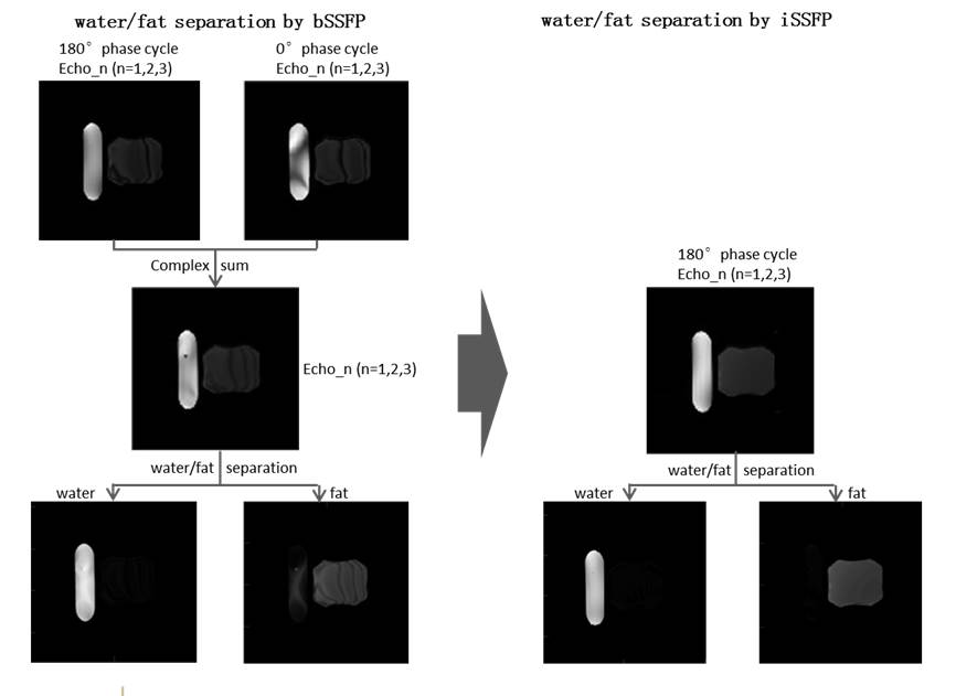
This study was performed at a 7T research system (Siemens, Erlangen, Germany) with a volume transmit/32 channel receive head coil (Nova Medical). The study was approved by the Institutional Review Board of Beijing MRI Center for Brain Research. The 3D iSSFP sequence is modified from 3D bSSFP by adding an extra dephasing gradient to cause a 2П phase dispersion across the width of a voxel along the readout orientation within a TR period2. The magnitude of the iSSFP signal was then kept constant irrespective of frequency shift. The chemical shift difference between water and fat is about 1040Hz at 7T. Three images of 3D iSSFP with different echo shifts were attained. Water and oil phantom experiment was performed to validate the behavior of water-fat decomposition for 3D iSSFP with the following parameters: Flip angle=30°,field of view=192*192mm2, number of slices=32, spatial resolution=1.15*1.15*3mm3, echo time=3.42/3.75/4.08ms, repetition time=7.08ms, scan time=2:15minutes. Images with different echo times were scanned in turn, respectively. 3D iSSFP images of foot were acquired from a healthy volunteer: Flip angle=30°,field of view=160*160mm2, number of slices=160, spatial resolution=0.6*0.6*0.6mm3, echo time=3.42/3.75/4.08ms, repetition time=7.08ms, GRAPPA acceleration factor=2, scan time=6:45minutes. As a reference, 3D bSSFP images for both 0°and 180°phase-cycling schemes were attained followed by a complex sum combination. Water and fat separation was performed with an efficient graph cut algorithm3.Results
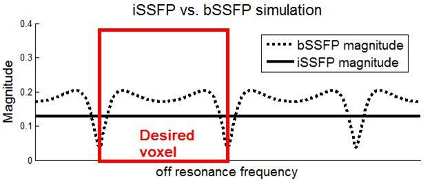
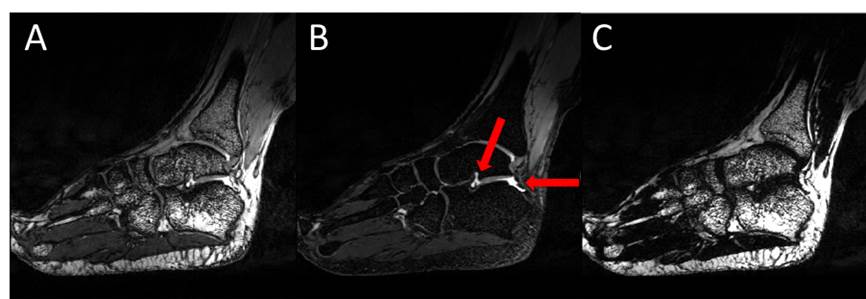
Bloch simulation demonstrated that iSSFP signal has constant magnitude across the frequency range of one cycle relative to bSSFP, thus showing no sensitivity to the banding artifacts (Fig. 1). 3D iSSFP and bSSFP with optimized echo times has been performed on the phantom and one healthy control. As shown in Fig. 2, bSSFP images still suffered from relieved banding artifacts, although the complex sum was performed. In comparison, 3D iSSFP produced better water/fat separation results with no banding artifacts using shorter scan time. The 7T in vivo experiment on human foot delineated the characteristics of iSSFP. In comparison with bSSFP (Fig. 3), iSSFP successfully separated water and fat, eliminated the banding artifacts and retained the advantages of high SNR and bright fluid (Fig. 4).Discussions and Conclusions
In this work, we have introduced a new water-fat separation technique known as iSSFP, which was modified from bSSFP. The iSSFP sequence inherited the characteristics of relatively high SNR and bright fluid. Simulation and in vivo experiments showed that 3D iSSFP was insensitive to field inhomogeneity and could produce better separation of water and fat in shorter scan time. 3D iSSFP with high spatial resolution may have great potential to separate the water and fat in the clinical application of 7T ultrahigh-field MRI.Acknowledgements
This work was supported in part by the Ministry of Science and Technology of China (MOST) grants (2015CB351701, 2012CB825500), National Nature Science Foundation of China grant (91132302), and Chinese Academy of Sciences Strategic Priority Research Program B grants (XDB02010001, XDB02050001).References
1. Quist B, Hargreaves BA, Daniel BL, Saranathan M. Balanced SSFP Dixon imaging with banding-artifact reduction at 3 Tesla. Magn. Reson. Med. 2015;74:706–715. doi: 10.1002/mrm.25449.
2. Sun K, Xue R, Zhang P, et al. Integrated SSFP for functional brain mapping at 7T with reduced susceptibility artifact. J. Magn. Reson. [Internet] 2017;276:22–30. doi: 10.1016/j.jmr.2016.12.012.
3. Berglund J, Kullberg J. Three-dimensional water/fat separation and T 2 * estimation based on whole-image optimization-Application in breathhold liver imaging at 1.5 T. Magn. Reson. Med. 2012;67:1684–1693. doi: 10.1002/mrm.23185.
Figures