5166
Cortical Bone Imaging Using Ultrashort Echo Time (UTE) Phase Sensitive Dual Inversion Recovery Subtraction Technique1University of California, San Diego, San Diego, CA, United States
Synopsis
Imaging of cortical bone is of fundamental importance in clinical MR but signals are low even with ultrashort echo time (UTE) sequences and contamination with high signals from long T2 muscle and fat is a problem 1. Here we propose a new phase sensitive dual inversion recovery subtraction method to obtain pure cortical bone images with zero signal from surrounding long T2 muscle and fat.
Introduction
Imaging of cortical bone is of fundamental importance in clinical MR but signals are low even with ultrashort echo time (UTE) sequences and contamination with high signals from long T2 muscle and fat can be problematic1. Here we propose a new phase sensitive dual inversion recovery subtraction method to obtain pure cortical bone images with zero signal from surrounding long T2 muscle and fat.Method
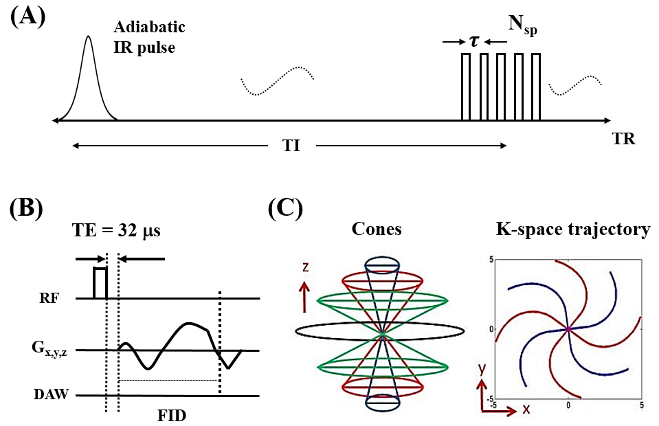
Features of the 3D adiabatic IR prepared UTE-Cones pulse sequence used in this study are shown in Figure 1 2. Following the IR preparation Nsp separate k-space spokes are acquired with an equal time interval τ between them to allow fast data acquisition. A short rectangular pulse (duration 26 to 52 µs) was used for non-selective signal excitation with each spoke (Fig. 1B), followed by 3D spiral trajectories with conical view ordering (Fig. 1C).
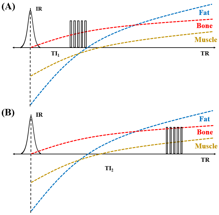
The signal variation during an IR UTE-Cones acquisition for bone, fat and muscle is shown in Figure 2. Bone signals are always positive due to the incomplete inversion of short T2 tissues. Fat and muscle signals are negative when using a short TI1 (Figs. 2A) but are positive when using a longer TI2 (Figs. 2B).
Two tibial datasets were acquired from a volunteer at two different TIs (TI = 100/250ms, TR = 300ms, flip angle = 18°, FOV=13×13×20cm, matrix=192×192×40, Nsp = 5) on a clinical 3T MRI scanner with an eight-channel knee coil for both signal transmission and reception.
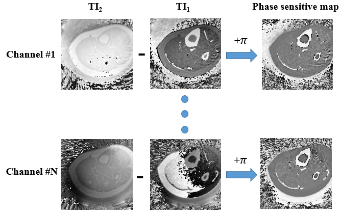
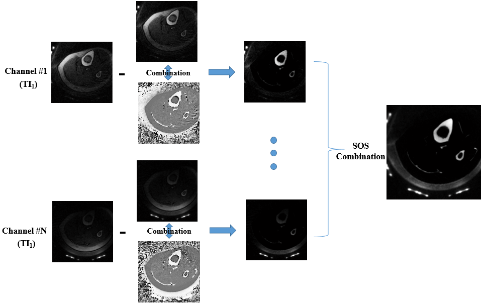
To obtain a pure phase map with polarized values for the inversion pulse, the background phases (from off-resonance induced phase shift and coil related phase offset) need to be removed first. As can be seen from Figure 3, such a phase sensitive map can be obtained by phase subtraction between the two datasets. A complex image can then be formed by combining the phase sensitive map and the original magnitude image. A pure bone image can be obtained by subtraction of the complex image from the original magnitude image (Figure 4). The final bone image is then obtained by sum of squares (SOS) combination of the eight channel bone images. Figure 4 shows the bone images from the data with TI1. Bone images at TI2 can also be produced using the same data processing procedure.
Results and Discussion
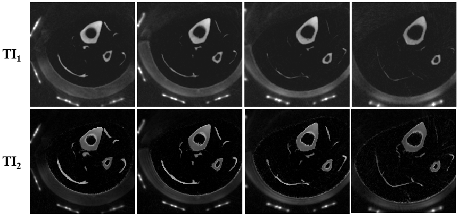
Figure 5 shows selected cortical bone maps generated using the proposed method. Two sets of bone images corresponding to TI1 and TI2 are shown.
The main reason to acquire the longer TI data is to obtain positive phase signal for all tissues. Other sequences, such as conventional 3D UTE-Cones sequence without preparation can also be used for this purpose.
Conclusion
The proposed phase sensitive dual inversion recovery subtraction method appears to a be very promising addition to bone MR imaging.Acknowledgements
The authors acknowledge grant support from NIH (1R01 AR062581 and 1R01 AR068987), VA Clinical Science Research & Development Service (1I01CX001388), and GE Healthcare.References
1. Du J, Bydder GM. Qualitative and quantitative ultrashort-TE MRI of cortical bone. NMR Biomed 2013;26:489-506.
2. Carl M, Bydder GM, Du J. UTE imaging with simultaneous water and fat signal suppression using a time-efficient multispoke inversion recovery pulse sequence. Magn Reson Med 2016;76:577–582.
Figures