5161
Imaging Scoliosis Using Zero Echo Time MRI1GE Healthcare, Taipei, Taiwan, 2Clinical Evoked Potential Study Room, Department of Orthopaedics and Traumatology, Taipei Veterans General Hospital, Taipei, Taiwan, 3Department of Radiology, Taipei Veterans General Hospital, Taipei, Taiwan, 4Division of Pediatric Orthopedics, Department of Orthopaedics and Traumatology, Taipei Veterans General Hospital, Taipei, Taiwan, 5Institute of Biomedical Engineering, National Yang Ming University, Taipei, Taiwan, 6Medicine, National Defense of Medical Center, Taipei, Taiwan
Synopsis
A high resolution, rapid scanning and three-dimensional zero-echo time (ZTE) protocol for cortical bone of whole spine imaging was established in this study. It provides computed tomography-like bone contrast and offers the similar result in the measurement of Cobb angle with conventional radiography, suggesting that the detection scoliosis and measurement of curvature using ZTE is possible, providing a potential alternative radiation-free diagnostic option that is especially relevant to scoliosis patients who need to undergo repeated examinations for evaluating the progress of spinal curvature.
Purpose:
Scoliosis, a three-dimensional (3D) deformity of the spinal column, is generally characterized by a lateral deviation of the spine, accompanied by an axial rotation of the vertebrae1. Radiography, computed tomography (CT), and magnetic resonance imaging (MRI) all have been used to play important roles in evaluating scoliosis and determining its underlying cause2,3. In contrast to the other two imaging modalities, MRI is capable of offering excellent soft-tissue detail about the nature of the spinal canal, cord and nerve root but fails to provide direct visualization of cortical bone due to its low water content and short signal life times (T2* ~ 0.4 ms), yielding limited cortical bone signal. A novel MRI technique named zero-echo time (ZTE) has been recent developed4. It utilizes hard pulse excitation followed by immediately 3D radial sampling to achieve nominal TE of zero and consequently could provide efficient sampling short T2* signals in cortical bone. The aim of this study is to first establish a clinical feasible ZTE whole spine protocol and assess ZTE imaging performance to scoliosis.Methods:
All MRI acquisitions were performed on a 1.5 T clinical scanner (Optima MR450w, GE Healthcare, Milwaukee, USA) using a large-size 16-channel flexible array coil as the signal detection and whole body coil for radio-frequency excitation. ZTE data were carried out in a prone position and acquired using a non-selective hard pulse excitation followed by 3D center-out radial sampling. The scanning parameters were as follows: TR=838 ms, flip angle=2 degree, receiver bandwidth= 31.2 kHz, field-of-view= 35 cm, isotropic resolution= 2.2 mm (interpolated to 1.36 × 1.36 × 1.1 mm), scan time= 2 min 20 s. Three-dimensional view of whole spine imaging was obtained on an Advantage Windows workstation using an advanced visualization platform (READY View (ver.4.6), GE Healthcare). The signal intensity of proton density-weighted ZTE images was inverted and rescaled to generate CT-like images proportional to tissue density.Results and Discussions:
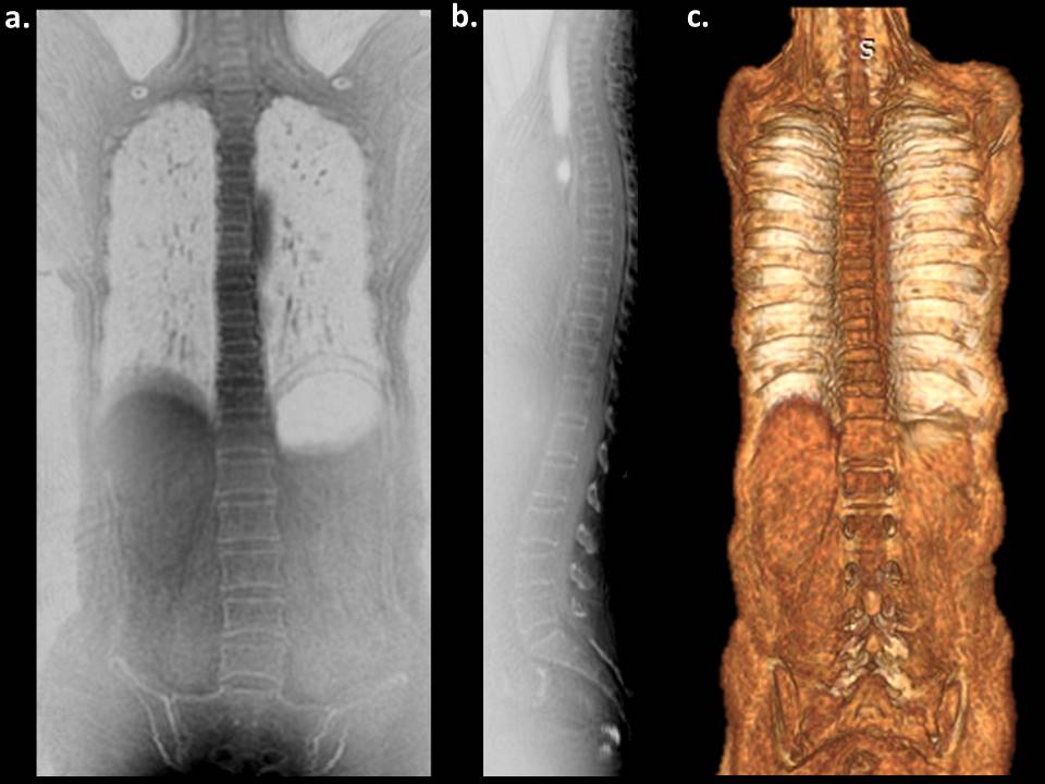
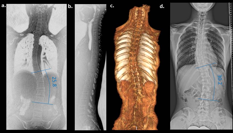
Whole spine ZTE MRI of a representative volunteer was shown in Fig. 1. Vertebral cortical bone was clearly visible at both views of ZTE images and there is no or minimal spine curvature to be visualized. Spinal curvature observed in the scoliosis patient by ZTE is similar with that in anteroposterior (AP) radiograph (Fig. 2). Whole spine ZTE imaging showed a coronal Cobb angle (T11-L3) of 25.8°, while the Cobb angle (T11-L3) measured at radiograph is 30.2°. As expected, the slight smaller Cobb angle was found at coronal ZTE image because MRI examination was performed in prone position, compared to AP radiograph in standing position. The ZTE performance in visualizing spinal cortical bone was consistence with both volunteer and scoliosis patient.Conclusion:
The preliminary results obtained in this study suggest that the detection scoliosis and measurement of curvature using ZTE is possible, providing a potential alternative radiation-free diagnostic option that is especially relevant to scoliosis patients who need to undergo repeated examinations for evaluating the progress of spinal curvature. Further assessment of more scoliosis patients with various level of curvature is currently underway.Acknowledgements
No acknowledgement found.References
1. Kim, Y, et al., “Scoliosis Imaging: What Radiologists Should Know,” RadioGraphs, 30:1823-42, 2010.
2. Cassar-Pullicino, VN, et al., “Imaging in scoliosis: what, why and how?” Clin Radiol, 57:543-562, 2002.
3. Schmitz, A, et al., “A new MRI technique for imaging scoliosis in the sagittal plane,” Eur Spine J, 10:114-117, 2001.
Figures