5152
Pressure-Triggered Gated MRI Acquisition of a Vibrating Scaled Vocal Fold Model1Electrical Engineering, Brigham Young University, Provo, UT, United States, 2Mechanical Engineering, Brigham Young University, Provo, UT, United States, 3Siemens Medical Solutions USA, Inc., Malvern, PA, United States
Synopsis
In this work, we present a technique for MR gated imaging of a scaled model of the vocal folds vibrating in the 13-17 Hz frequency range. The vocal fold model was scaled to roughly 4X human size to decrease the frequency of vibration and make gating feasible. The model contained an embedded grid of markers such that biomechanical motion of the laryngeal model could be accurate tracked across multiple phases of the periodic vibrational cycle.
Purpose
MRI of periodically moving objects often utilizes physiological signals, such as ECG, to gate cardiac images in CINE imaging1 or respiratory gating to eliminate motion in the abdominal and thorax regions2. However, when the period of motion becomes too short, gating techniques are much more challenging. There is significant interest in imaging of the larynx and vocal folds for a variety of applications3-4, but the period of vibration in the vocal folds (typically <10ms) is such that gated imaging has been impractical. This rapid vocal fold motion, while periodic, complicates acquisition of images and results in significant motion blurring.Vocal fold tissue engineering and the development of biomaterials for treatment of voice disorders can be aided by an understanding of the internal stress and strain field experienced by the vocal folds during phonation. While conventional imaging (e.g., using high-speed cameras) has been used to image the exterior surfaces of synthetic replicas during vibration, imaging of the internal regions has not been possible due to limited optical access. In this study we show that gated MRI can be used for internal imaging and deformation tracking through the use of an embedded grid of markers in a scaled-up (4x) synthetic vocal fold model.We created a 4x scale synthetic vocal fold model that resonates at a much lower frequency (~15 Hz) compared to human vocal folds (>100 Hz). At this frequency, gated MR imaging becomes practical. An MRI protocol was developed for imaging the cross section of these scaled-up synthetic vocal fold replicas during vibration. We demonstrate a method of effectively freezing the motion at sequential phases through use of the pressure wave generated by the movement of the scaled model to trigger the acquisition of a phase encode line in an acquired image. Analysis of the motion within these larger scale models will allow validation of computational models of vocal fold motion.Methods
Apparatus
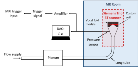
The vocal fold models are fixed in a shroud. Tubing extends from the bottom of the shroud and connects to an air supply. When the subglottal pressure reaches 0.15 - 0.17 kPa, the model begins to vibrate. The vibration frequency ranges from 13 to 16 Hz with amplitudes of 7 - 8.5 mm. The experimental setup can be seen in Figure 1. The experimental apparatus inside the MRI consists of the 3D printed shroud and the synthetic vocal folds. A custom 6 inch single loop hydrogen coil was constructed to fit directly onto the experimental apparatus instead of using the body coil to achieve higher SNR. Subglottal tubing extended outside of the MRI room and connected to the air source. Subglottal pressure and frequency were recorded via a data acquisition system connected to a pressure sensor located in the subglottal region. The subglottal pressure signal was amplified, conditioned, and fed into the MRI as the external input on the physio card.
MRI Protocol
MRI images were acquired on a Siemens MAGNETOM Tim Trio (Siemens Healthineers, Elrangen Germany) using a GRE sequence triggered by the external input on the physio card. High resolution 2D images were acquired using a 128x128 mm FOV, 256x256 matrix size, 4mm slice thickness, TE=2.7 ms, TR as determined by the external trigger, approximately 65 ms, flip angle=10°, 3/4 partial Fourier phase acquisition, bandwidth=590 Hz/pixel, and 10 averages. High resolution 3D volume scans were acquired using a 128x128 mm FOV, 1 mm slice thickness, 128x128x72 matrix size, TE=2.1 ms, T2 as determined by the external trigger input, approximately 65 ms, flip angle=10°, 3/4 slice and 3/4 phase partial Fourier acquisition, and a bandwidth=797 Hz/pixel. In both cases, a trigger delay was used to capture the phantom motion at different phases of its movement.
Results
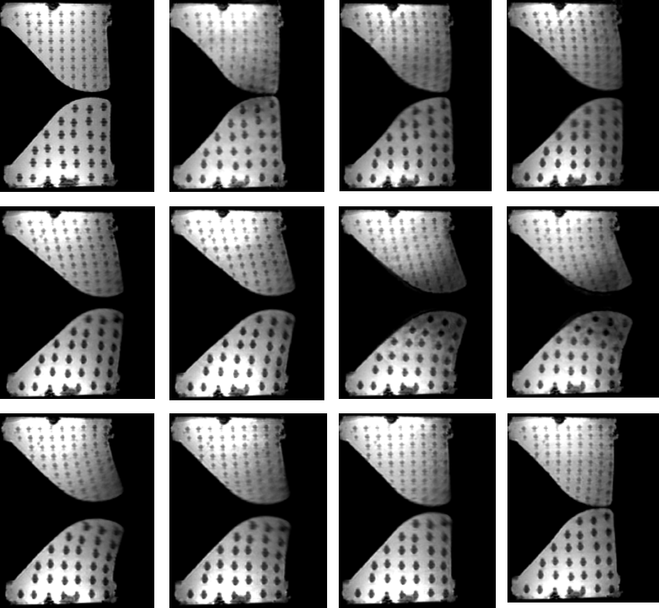
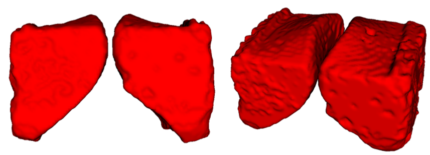
We demonstrated the ability to capture multiple phases of motion in a 4X scale model of the vocal folds using external triggering. Motion tracking was sufficient to allow averaged 2D and 3D scans without significant motion blurring. Results from 2D scans are shown in Figure 2 with a complete cycle of phases. Despite some blurring of the tracking grid, center points can be registered and tracked to measure the internal motion of the model. An entire 3D volume of the phantom in motion is rendered in Figure 3. This allows tracking the motion of the model surface.Discussion
The work presented demonstrates the capability to reproduce images of an oscillating vocal fold model, allowing motion to be frozen at any arbitrary phase using either 2D or 3D acquisitions. Future work will explore the possibility of imaging smaller scale models, and potentially performing similar in-vivo studies as gradient capabilities increase allowing shorter readout durations and gating of higher-frequency vibration.Acknowledgements
We would like to acknowledge funding from National Institutes of Health 2 R56 DC009616-06.References
1. Atkinson D.J., Edelman R.R. Cineangiography of the heart in a single breath hold with a segmented TurboFLASH sequence. Radiology 1991; 178:357-360.
2. Ehman R.L., McNamara M.T., et al. Magnetic resonance imaging with respiratory gating: techniques and advantages. Am J Roentgenol. 1984;143(6):1175-82.
3. Ahmad M., Dargaud J., et al. Dynamic MRI of Larynx and Vocal Fold Vibrations in Normal Phonation. Journal of Voice 2009; 23(2):235-239.
4. Baki, M.M., Menys, A., et al. Feasibility of vocal fold abduction and adduction assessment using cine-MRI. Eur Radiol 2107; 27: 598.
Figures