5149
MAVRIC SL Compressed Sensing STIR Imaging using a Total Generalized Variation (TGV) Reconstruction1MR Applications and Workflow, GE Healthcare, Waukesha, WI, United States, 2Hospital for Special Surgery, New York City, NY, United States, 3Medical College of Wisconsin, Milwaukee, WI, United States
Synopsis
3D Multi-spectral imaging sequences like MAVRIC SL have been used overcome susceptibility artifacts caused by metallic hardware. Several studies have attempted to reduce long scan times by using parallel imaging and compressed sensing, but PD images are commonly displayed with evident blurring. MAVRIC STIR images, due to their sparseness, may be more amenable to compressed sensing acceleration. The current work focuses on accelerating MAVRIC SL STIR images using compressed sensing and a total generalized variation reconstruction (TGV).
Introduction
3D Multi-Spectral Imaging sequences, like MAVRIC SL and SEMAC, significantly reduce the susceptibility artifacts caused by metal instrumentation [1,2]. This artifact reduction is achieved by sampling the broadened frequency distribution and acquiring images (spectral bins) at distinct frequency offsets from the Larmor frequency, which are combined to minimize the susceptibility artifacts. The longer scan times of these series have been accelerated using parallel imaging, partial Fourier acquisition, and cutting the corners of k-Space. In addition, compressed sensing has been used in combination with parallel imaging to accelerate the 3D MSI acquisition [3,4,5]. Koch et al. showed that wavelet based regularization in the ARC+CS algorithm leads to prohibitive blurring in PD images [3]. More recently, Levine et al. used a Low Rank + sparse algorithm – which used in-plane acceleration and further accelerated the bin dimension using a variable density sampling pattern – to significantly reduce scan time [5]. While this resolved blurring away from the metal, regions closer to the implant were still affected. In this project, we focus the compressed sensing reconstruction on the sparser STIR images, which should more readily lend itself to compressed sensing acceleration. Here, we apply a total generalized variation (TGV) reconstruction [6] to reconstruct individual spectral-bins before they are combined to yield an artifact minimized image.Methods
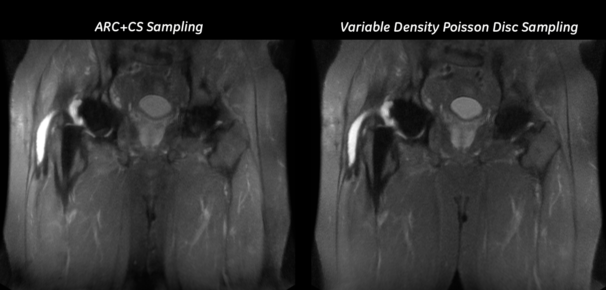
Images were acquired on a clinical 1.5T MR450W scanner (GE Healthcare, Waukesha WI), under an IRB approved study with written informed consent from the patients. A spectral calibration scan was run to determine the number of bins needed for each implant [7]. A conventional MAVRIC SL STIR was acquired with 24 spectral bins and the following parameters: matrix: 256x192, FOV: 20 – 40cm, slice thickness: 5.5mm, ETL: 20, TE/TI/TR: 7/150/4237ms, NEX: 0.5, ARC: 2x2. The STIR acquisition was repeated with a B1-optimized inversion pulse [8], fewer spectral bins (as determined by the calibration scan), and the following parameters: matrix: 256x192, FOV: 20 – 43cm, slice thickness: 4mm-5.5mm, ETL: 48 with variable flip angles, TE/TI/TR: 7/150/4237ms, NEX: 1, ARC: 2x2, CS factor: 1.4. To test the impact of the sampling pattern on image quality, in one subject, higher resolution images were acquired using both the ARC+CS sampling pattern and a variable density Poisson disc sampling (VDPDS). The images were reconstructed first using the ARC+CS algorithm which first performs wavelet regularized reconstruction using a conjugate gradient descent optimizer to fill the randomly undersampled locations, and then performs ARC reconstruction to fill the uniformly under sampled locations. The data were also reconstructed using the TGV algorithm which iteratively fills in both the regularly and randomly undersampled regions of k-space. The coil sensitivity profiles were determined by low pass filtering the k-space data, the TGV regularization constant was empirically chosen to be 0.5, and the algorithm used 50 iterations for each spectral bin. Reconstruction was done in C++ using the Orchestra SDK (GE Healthcare, Waukesha WI).Results
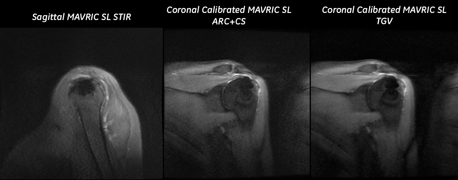
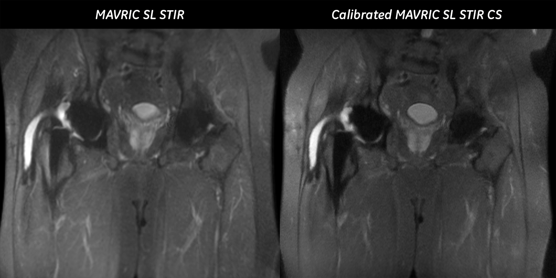
Figure 1 shows data from a subject with an implanted suture anchor to secure the supraspinatus tendon to the proximal humerus. This data set was acquired with 8 spectral bins, in 3:30 min. The conventional ARC+CS reconstruction retains a significant degree of noise, which has been minimized with the TGV reconstruction. An additional example of the improvement to the STIR image quality can be seen in Figure 2, where the lower resolution of the product STIR could be traded in for higher resolution, a full-Fourier acquisition, thinner slices, and with the addition of compressed sensing, can be achieved in a scan time that is on par with the lower resolution clinical scan (4:23 min vs. 5:11 min). Different sampling patterns are compared in figure 3, with the VDPDS sampling resulting in a more uniform image.Discussion and Conclusions
For the sparse MAVRIC SL STIR images, the TGV regularizer is advantageous as it does not severely penalize edges and also denoises the image. This features permits reduction of the voxel size with a concomitant full-Fourier acquisition to negate the SNR losses induced by a homodyne reconstruction. Most importantly, these features can be implemented while maintaining a scan time similar to the lower resolution clinical STIR acquisition. The data set acquired with a variable density Poisson disc sampling suggests that a choice of sampling pattern may potentially improve the performance of the TGV reconstruction.Acknowledgements
No acknowledgement found.References
1. Koch, KM, Brau, AC, Chen, W et al. “Imaging near metal with a MAVRIC-SEMAC hybrid”, Magn Reson Med 2011;65:71-82.
2. Lu, Wenmiao, et al. "SEMAC: slice encoding for metal artifact correction in MRI." Magnetic resonance in medicine 62.1 (2009): 66-76.
3. Koch, KM, King, KF “Combined parallel imaging and Compressed Sensing on 3D Multi Spectral Imaging near metal implants”, #3172, Proceedings of the ISMRM 2011.
4. Worters, Pauline W., et al. "Compressed‐Sensing multispectral imaging of the postoperative spine." Journal of Magnetic Resonance Imaging 37.1 (2013): 243-248.
5. Levine, Evan, et al. "Accelerated three‐dimensional multispectral MRI with robust principal component analysis for separation of on‐and off‐resonance signals." Magnetic Resonance in Medicine (2017).
6. Knoll, Florian, et al. "Second order total generalized variation (TGV) for MRI." Magnetic resonance in medicine 65.2 (2011): 480-491.
7. Kaushik, S.S. et al., "External calibration of the spectral coverage for three‐dimensional multispectral MRI." Magnetic resonance in medicine 76.5 (2016): 1494-1503.
8. Kaushik, S.S. et al., “Lipid suppression around metal implants using a B1-optimized adiabatic inversion pulse”, #5028, Proceedings of the ISMRM 2016.
Figures