5147
Can 5-Minute 3D Isotropic Turbo Spin-Echo MR Imaging Replace 2D Standard Knee MR Imaging at 3T?1Radiology, Seoul St. Mary’s Hospital, College of Medicine, The Catholic University of Korea, Seoul, Republic of Korea, 2Radiology, Seoul St. Mary’s Hospital, College of Medicine, The Catholic University of Korea, Seocho-gu, Republic of Korea, 3Occupational and environmental medicine, Seoul St. Mary’s Hospital, College of Medicine, The Catholic University of Korea, Seocho-gu, Republic of Korea, 4Orthopedic surgery, Seoul St. Mary’s Hospital, College of Medicine, The Catholic University of Korea, Seoul, Republic of Korea, 5Radiology, Seoul St. Mary's Hospital, The Catholic University of Korea, Seoul, Republic of Korea
Synopsis
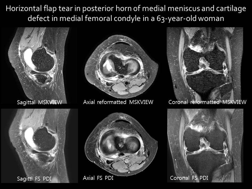
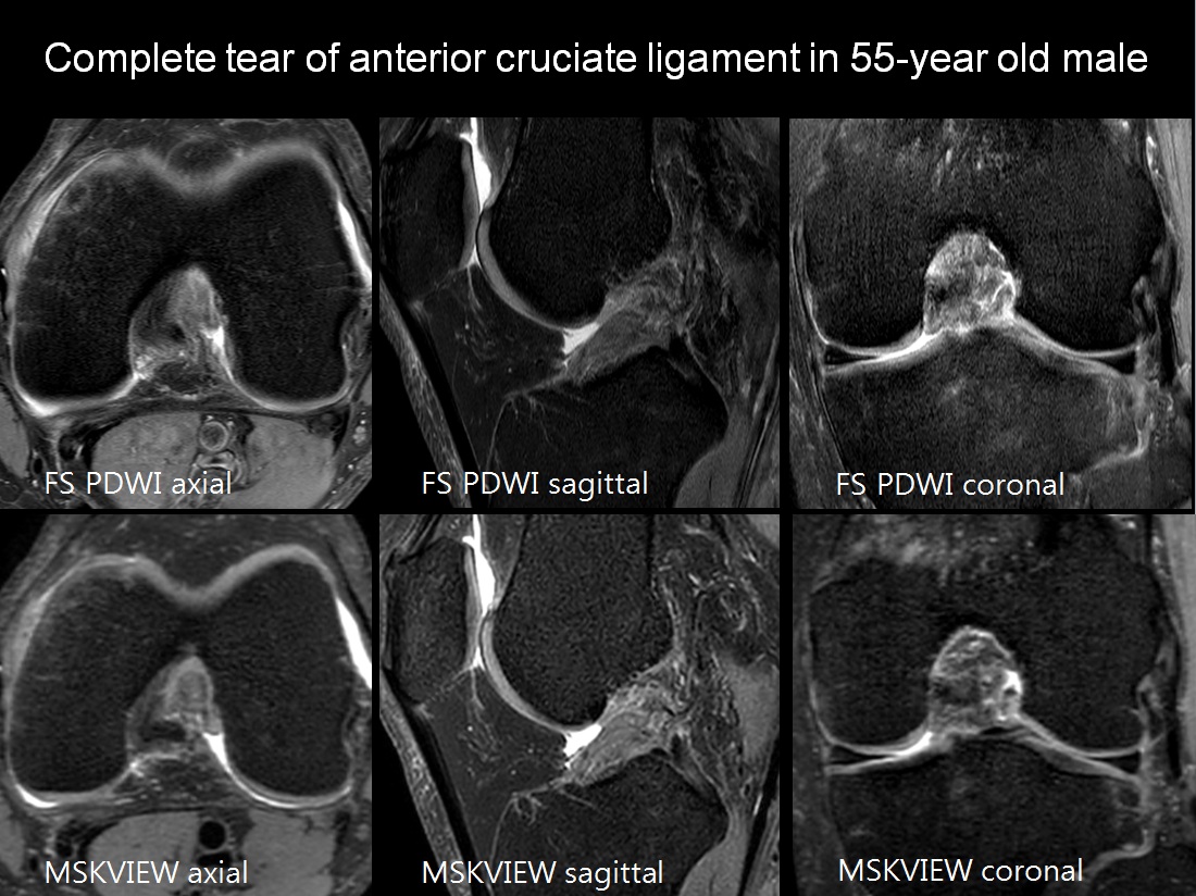
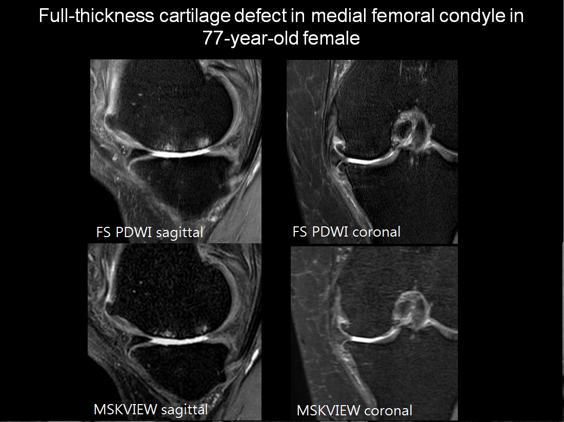
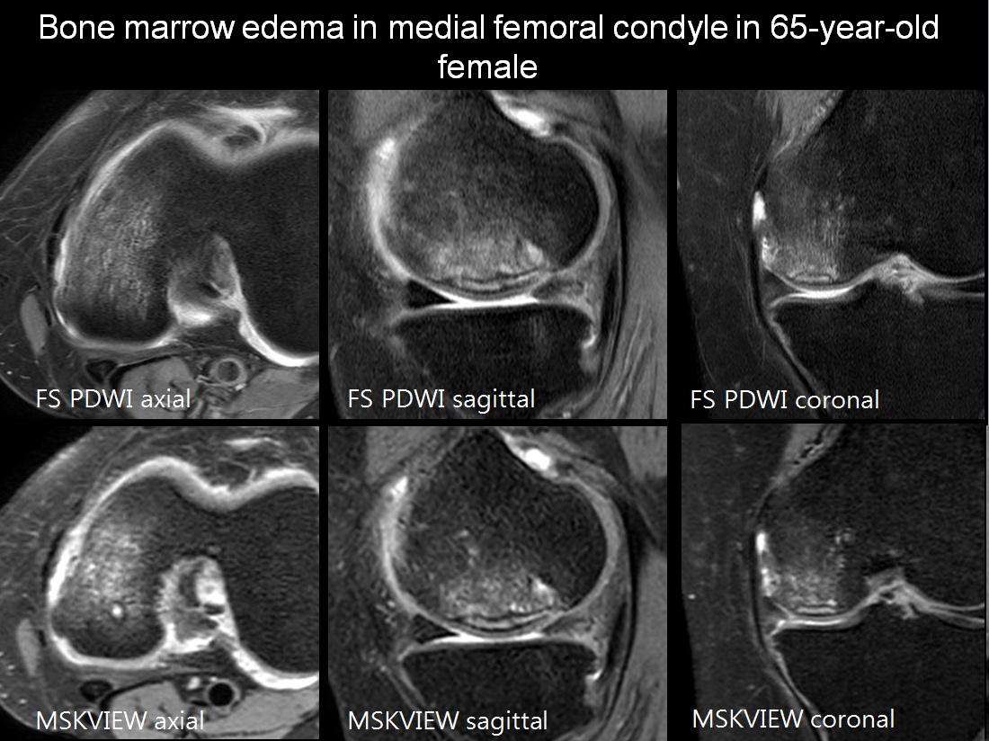
2D TSE intermediate(IM) or T2-weighted images have been traditionally used to evaluate internal derangements of knee. However partial volume effect due to relatively thick image section and gap and long acquisition time due to impossible reformation is limitation of standard 2D TSE technique. Newly developed 3D isotropic T2 or IM weighted image is made by isotropic voxel data, which allows 3D reformation and reduce acquisition time. The purpose of this study is to compare the image quality and diagnostic performance of 5-minute sagittal fat-suppressed 3D isotropic turbo spin-echo sequence (MSK VIEW) and 2D standard knee magnetic resonance (MR) imaging at 3T.
Purpose
2D TSE intermediate(IM) or T2-weighted images have been traditionally used to evaluate internal derangements of knee. However partial volume effect due to relatively thick image section and gap and long acquisition time due to impossible reformation is limitation of standard 2D TSE technique. Newly developed 3D isotropic T2 or IM weighted image is made by isotropic voxel data, which allows 3D reformation and reduce acquisition time. The purpose of this study is to compare the image quality and diagnostic performance of 5-minute sagittal fat-suppressed 3D isotropic turbo spin-echo sequence (MSK VIEW) and 2D standard knee magnetic resonance (MR) imaging at 3T.
Materials & Methods
Subjects
The institutional review board approved this retrospective study and informed consent was waived. Fifty consecutive patients (mean age, 57.4 ± 11.2; range, 25-78 years) who underwent 3T knee MR imaging with 5-minute MSKVIEW and SPIR between September 2016 and January 2017 were included. Patients with history of prior knee surgery were excluded.
MRI protocol
All patients were scanned on a Philips 3T MRI system. Each patients were scanned MRI including these sequences: Sagittal MSKVIEW with coronal and axial reformatted images; SPIR with axial, coronal, sagittal plane
Image analysis
Three orthogonal planes were separately reviewed by two musculoskeletal radiologists. Two data sets were reviewed with three weeks interval. Readers graded image quality using two level scales for seven criteria using published classification system: optimal or suboptimal for edge sharpness, blurring, artifact, contrast between fluid and cartilage, small ligament delineation, and amount of noise. Readers detected lesions using five-level confidence score for the presence of medial meniscal tears, lateral meniscal tears, anterior cruciate ligament (ACL), posterior cruciate ligament (PCL), medial collateral ligament (MCL), and lateral collateral ligament (LCL) tears, and the presence of cartilage abnormality in medial femoral condyle, and bone marrow edema. Differences in image quality between MSKVIEW and SPIR was assesed using Student's t-test, respectively. Interprotocol agreement between MSKVIEW and SPIR for each lesion was assessed using weighted к in two readers, respectively.
Results
There were no significant differences in image quality between two protocols, except for motion artifact in SPIR image in four cases (p<0.0001). SPIR showed motion artifact due to popliteal arterial pulse in all patients, whereas none of MSKVIEW did not show motion artifact. Interprotocol agreements were substantial to very good for medial meniscus and excellent in lateral meniscus, all ligaments, and articular cartilage of medial femoral condyle: κ=0.728 and κ=0.954 for medial meniscal tears; κ=0.935 and κ=0.922 for lateral meniscal tears; κ=0.821 and κ=0.83 for ACL tears; κ=0.871 and κ=0.903 for PCL tears; κ=0.929 and κ=0.851 for MCL tears; κ=0.82 and κ=0.9 for LCL tears; κ=0.925 and κ=0.844 for articular cartilage of medial femoral condyle; and κ=0.973 and κ=0.932 for bone marrow edema in reader 1 and reader 2, respectively.Conclusion
Five-minute sagittal fat-suppressed 3D isotropic turbo spin-echo sequence may be replaceable for 2D standard knee MR imaging at 3T.Acknowledgements
No acknowlegement found.References
1. Escobedo EM, Hunter JC, Zink-Brody GC, Wilson AJ, Harrison SD, Fisher DJ (1996) Usefulness of turbo spin-echo MR imaging in the evaluation of meniscal tears: comparison with a conventional spin-echo sequence. American Journal of Roentgenology 167:1223-1227 2
2. Jee W-H, McCauley TR, Kim J-M et al (2003) Meniscal Tear Configurations: Categorization with MR Imaging. American Journal of Roentgenology 180:93-97 3
3. Schaefer FK, Schaefer PJ, Brossmann J et al (2006) Value of fat-suppressed PD-weighted TSE-sequences for detection of anterior and posterior cruciate ligament lesions--comparison to arthroscopy. Eur J Radiol 58:411-415 4
4. Sonin AH, Pensy RA, Mulligan ME, Hatem S (2002) Grading Articular Cartilage of the Knee Using Fast Spin-Echo Proton Density-Weighted MR Imaging Without Fat Suppression. American Journal of Roentgenology 179:1159-1166 5
5. Bredella MA, Tirman PF, Peterfy CG et al (1999) Accuracy of T2-weighted fast spin-echo MR imaging with fat saturation in detecting cartilage defects in the knee: comparison with arthroscopy in 130 patients. AJR Am J Roentgenol 172:1073-1080
Figures