5142
Reproducibility of MR elastography of lumbar paraspinal muscle in healthy volunteers1Department of Medical Imaging and Radiological Science, Central Taiwan University of Science and Technology, Taichung, Taiwan, 2Department of Radiology, National Cheng Kung University Hospital, Tainan, Taiwan
Synopsis
The abnormal tissue stiffness of lumbar paraspinal muscle in symptomatic patients can be diagnosed by palpation in clinical practice. MRE can measure tissue stiffness quantitatively and noninvasively. The objective of this pilot study was to evaluate the reproducibility of MRE of lumbar paraspinal muscles in healthy volunteers. The strong positive relationship between 2 MRE exams was demonstrated by the Pearson's correlation coefficient.
Purpose
To evaluate the reproducibility of MR elastography of lumbar paraspinal muscle in healthy volunteers.Introduction
Magnetic resonance elastography (MRE) is a non-invasive MR-based phase contrast imaging technique that applied an oscillating motion to detect tissue vibratory displacements. Wavelengths are then determined from the images and used to calculate the speed of the waves. Initial efforts were devoted to the application of the approach to soft tissues such as the breast and liver. More recent work has begun to assess its utility in the study of skeletal muscles in the upper and lower extremities1.
Lower back pain (LBP) is very common in clinical practice. One of the underlying mechanisms of LBP is the development of the myofascial trigger points that are usually accompanied by the palpable, tender nodules in taut bands2.
In this study, we will apply MRE examinations in the healthy individuals and investigate whether MRE is a valid and reproducible technique for objectively estimating tissue stiffness of lower back by 2 independent analysts.
Methods
Sixty healthy volunteers (35 men, 25 women, age range: 21-57 years) were enrolled in this MRE study. All MR images were obtained by using a 3-T MR scanner (Ingenia; Philips Medical Systems, Best, the Netherlands) with a SENSE Flex-M coil and a gradient system providing 80 mT/m and a slew rate of 200 mT/m/ms. Subjects laid prone with the relaxed arms along the both sided of the body to prevent movement during image acquisition. The MR-compatible transducer has been designed by the Philips Medical Systems with the vibration wave frequency range from 40 to 200 Hz. The transducer was positioned on the midline of the lower back. Two same-day MRE exams were separated by a 10-min break. Between exams, subjects were removed and then repositioned on the scanner table.
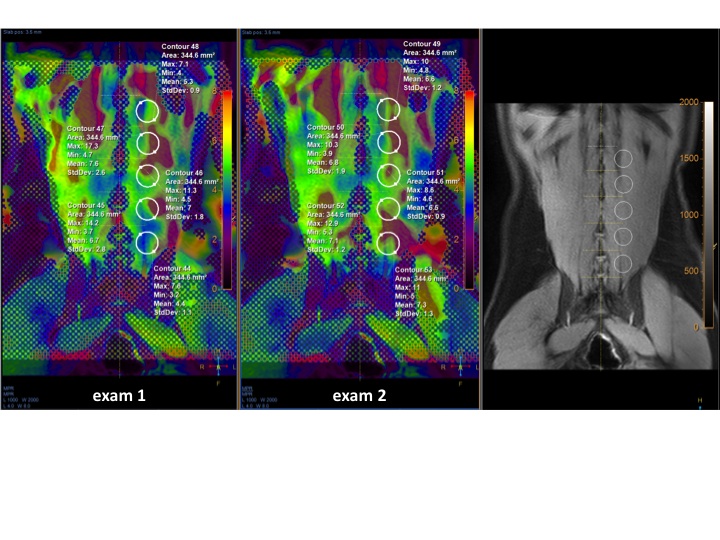
Two independent analysts measured the viscoelastic values of right or left paraspinal muscles on the 4 coronal images in different depth. The lateralization measurements were randomized. The circular regions of interest (ROIs) were carefully placed at the L1, L2, L3, L4, and L5 levels to avoid the regions including ineffective data . ROIs were co-localized automatically to the corresponding images in the second exam. Reproducibility between 2 exams was assessed by the Pearson's correlation coefficient.
Results
The areas of circular ROIs ranged from 193.7 mm2 to 443.6 mm2 by the analyst 1, and from 189.4 mm2 to 533.4 mm2 by the analyst 2. The ROIs were placed at the same side of the lower back on the color-coded elastogram (Fig) of the 2 MRE exams. The mean viscoelastic values of the paraspinal muscles were 6.67 ± 1.99 kPa for the first MRE exam, and 6.61 ± 2.00 kPa for the second MRE exam by the analyst 1, then the mean viscoelastic values were 6.70 ± 1.94 kPa for the first MRE exam, and 6.64 ± 1.94 kPa for the second MRE exam by the analyst 2. The Pearson's correlation coefficient between 2 MRE exams was 0.63 for analyst 1, and 0.64 for analyst 2.Conclusion
There was strong positive relationship between 2 MRE exams by 2 independent analysts in this study. Therefore, MRE is a valid and reproducible technique for objectively assessing tissue stiffness of lumbar paraspinal muscles.Acknowledgements
This study was supported by a grant from Ministry of Science and Technology, Taiwan, under the contract MOST 105-2314-B-006-047-.References
1. Green MA, Geng G, Qin E, Sinkus R, Gandevia SC, Bilston LE. Measuring anisotropic muscle stiffness properties using elastography. NMR Biomed 2013;26(11):1387-1394.
2. Chen Q, Bensamoun S, Basford JR, Thompson JM, An KN. Identification and quantification of myofascial taut bands with magnetic resonance elastography. Arch Phys Med Rehabil 2007;88(12):1658-1661.
Figures