5140
A static MR follow-up study of injured levator ani muscle recovery1radiology, Tianjin first center hospital, china, tianjin, china, China, 2tianjin first center hospital, tianjin, China, 3Philips Healthcare, Beijing, China
Synopsis
The abnormal structure or function of Levator ani muscle(LAM) is the basis for pelvic floor dysfunction disease. Our study is to assess the recovery of injured LAM resulting from vaginal delivery by using static MRI.The primiparas who presented LAM injury at six weeks after delivery were brought into MRI follow-up study(reviewed at three months and six months).54 pubovisceralis injury with edema in a bilateral summary, there was significant difference among different postpartum time points. To summarise, the injured LAM has the ability to recover after delivery. LAM edema may exaggerate the true severity and extent of LAM injury.
Objective
The abnormal morphology or function of Levator ani muscle(LAM) is the basis for pelvic floor dysfunction disease. Vaginal delivery is the most important risk factor resulting in LAM injury. However, we has little knowledge about the recovery of injured LAM after delivery, how much impact of the abnormal LAM structure defect and whether it needs pelvic intervention treatment. In addition, it is necessary to deeply understand how to choose the best imaging patterns for evaluating LAM injury and diagnostic considerations. So, our study is to assess the recovery of injured LAM resulting from vaginal delivery by using static MRI.Methods
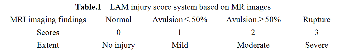
120 primiparas were prospectively collected and underwent static MRI imaging by using a 3T MR scanner (Ingenia, Philips Healthcare, the Netherlands) at the time point of six weeks after delivery.The scanning protocol was FSE T2WI sequence and scan parameters were as followed: TR=4236-4413 ms, TE=100 ms, FOV=260 mm×260 mm, thickness=3.0 mm, Slice Number 25. LAM injury were evaluated on T2WI images according to the LAM injury score system1 based on MR images(Table.1). The primiparas who presented LAM injury were brought into MRI follow-up study(reviewed at three months and six months). Two radiologists assessed LAM injury among all the subjects. The evaluation consistency of two radiologists were evaluated using kappa test. Chi-square test were used to compare severity distribution of injured LAM at different postpartum time.Results
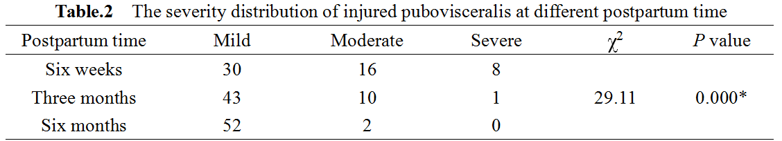
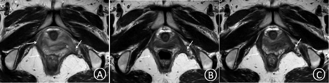
34 primiparas presented LAM injury among 120 cases, but there were a total of 54 pubovisceralis injury with edema in a bilateral summary(Fig.1) and the severity distribution at different postpartum time were listed in Table.2, there was significant difference among three groups (χ2=29.11, P=0.000). There were significant differences between six weeks and three months, six weeks and six months, three months and six months after compared in pairs(χ2=9.144, P=0.010; χ2=24.791, P=0.000: χ2=7.186, P=0.015).Discussion
In the follow-up study, the injured LAM scores during six weeks, three months and six months were decrease, reminding that the injured LAM has the ability to recover after delivery. Interestingly, LAM edema was obvious in the early stage after delivery and we can not finger out the continuity of muscle fibers, which will interfere with injury score2. LAM injury may be overdiagnostic as a result of edema early after delivery.Conclusion
LAM injury caused by vaginal delivery has a natural capacity of recovery, LAM edema may exaggerate the true severity and extent of LAM injury(avulsion, rupture) and obstruct diagnosticoverdiagnose assessment.
Acknowledgements
No acknowledgement found.References
1.Morgan DM, Umek W, Stein T, et al. Interrater reliability of assessing levator ani muscle defects with magnetic resonance images[J]. Int Urogynecol J Pelvic Floor Dysfunct, 2007, 18(7): 773-778.
2.Miller JM, Brandon C, Jacobson JA, et al. MRI findings in patients considered high risk for pelvic floor injury studied serially after vaginal childbirth. AJR Am J Roentgenol 2010;195:786-91.
Figures